7LC0

Structure of D-Glucosaminate-6-phosphate Ammonia-lyase


Experimental Data Snapshot

  • Method: X-RAY DIFFRACTION
  • Resolution: 2.60 Å
  • R-Value Free: 0.228 
  • R-Value Work: 0.193 
  • R-Value Observed: 0.194 

wwPDB Validation   3D Report Full Report


This is version 1.0 of the entry. See complete history


Literature

Structure and Mechanism of d-Glucosaminate-6-phosphate Ammonia-lyase: A Novel Octameric Assembly for a Pyridoxal 5'-Phosphate-Dependent Enzyme, and Unprecedented Stereochemical Inversion in the Elimination Reaction of a d-Amino Acid.

Phillips, R.S.Ting, S.C.Anderson, K.

(2021) Biochemistry 60: 1609-1618

  • DOI: https://doi.org/10.1021/acs.biochem.1c00106
  • Primary Citation of Related Structures:  
    7LC0, 7LCE

  • PubMed Abstract: 

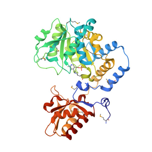
    d-Glucosaminate-6-phosphate ammonia-lyase (DGL) is a pyridoxal 5'-phosphate (PLP)-dependent enzyme that produces 2-keto-3-deoxygluconate 6-phosphate (KDG-6-P) in the metabolism of d-glucosaminic acid by Salmonella enterica serovar typhimurium. We have determined the crystal structure of DGL by SAD phasing with selenomethionine to a resolution of 2.58 Å. The sequence has very low identity with most other members of the aminotransferase (AT) superfamily. The structure forms an octameric assembly as a tetramer of dimers that has not been observed previously in the AT superfamily. PLP is covalently bound as a Schiff base to Lys-213 in the catalytic dimer at the interface of two monomers. The structure lacks the conserved arginine that binds the α-carboxylate of the substrate in most members of the AT superfamily. However, there is a cluster of arginines in the small domain that likely serves as a binding site for the phosphate of the substrate. The deamination reaction performed in D 2 O gives a KDG-6-P product stereospecifically deuterated at C3; thus, the mechanism must involve an enamine intermediate that is protonated by the enzyme before product release. Nuclear magnetic resonance (NMR) analysis demonstrates that the deuterium is located in the pro - R position in the product, showing that the elimination of water takes place with inversion of configuration at C3, which is unprecedented for a PLP-dependent dehydratase/deaminase. On the basis of the crystal structure and the NMR data, a reaction mechanism for DGL is proposed.


  • Organizational Affiliation

    Department of Chemistry, University of Georgia, Athens, Georgia 30602, United States.


Macromolecules
Find similar proteins by:  (by identity cutoff)  |  3D Structure
Entity ID: 1
MoleculeChains Sequence LengthOrganismDetailsImage
Putative selenocysteine synthase (L-seryl-tRNA(Ser) selenium transferase)
A, B, C, D, E
A, B, C, D, E, F, G, H
369Salmonella enterica subsp. enterica serovar Typhimurium str. LT2Mutation(s): 0 
Gene Names: STM3768
UniProt
Find proteins for Q8ZL27 (Salmonella typhimurium (strain LT2 / SGSC1412 / ATCC 700720))
Explore Q8ZL27 
Go to UniProtKB:  Q8ZL27
Entity Groups  
Sequence Clusters30% Identity50% Identity70% Identity90% Identity95% Identity100% Identity
UniProt GroupQ8ZL27
Sequence Annotations
Expand
  • Reference Sequence
Small Molecules
Ligands 5 Unique
IDChains Name / Formula / InChI Key2D Diagram3D Interactions
PEG
Query on PEG

Download Ideal Coordinates CCD File 
S [auth C]DI(HYDROXYETHYL)ETHER
C4 H10 O3
MTHSVFCYNBDYFN-UHFFFAOYSA-N
DMS
Query on DMS

Download Ideal Coordinates CCD File 
BA [auth E]
CA [auth E]
DA [auth E]
FA [auth F]
GA [auth F]
BA [auth E],
CA [auth E],
DA [auth E],
FA [auth F],
GA [auth F],
HA [auth F],
I [auth A],
IA [auth F],
K [auth A],
KA [auth G],
LA [auth G],
MA [auth G],
N [auth B],
O [auth B],
P [auth B],
T [auth C],
V [auth D],
W [auth D],
X [auth D]
DIMETHYL SULFOXIDE
C2 H6 O S
IAZDPXIOMUYVGZ-UHFFFAOYSA-N
ACY
Query on ACY

Download Ideal Coordinates CCD File 
AA [auth E],
J [auth A],
M [auth B],
Y [auth E],
Z [auth E]
ACETIC ACID
C2 H4 O2
QTBSBXVTEAMEQO-UHFFFAOYSA-N
CL
Query on CL

Download Ideal Coordinates CCD File 
R [auth B]CHLORIDE ION
Cl
VEXZGXHMUGYJMC-UHFFFAOYSA-M
MG
Query on MG

Download Ideal Coordinates CCD File 
EA [auth E]
JA [auth F]
L [auth A]
NA [auth G]
OA [auth G]
EA [auth E],
JA [auth F],
L [auth A],
NA [auth G],
OA [auth G],
PA [auth G],
Q [auth B],
QA [auth H],
RA [auth H],
SA [auth H],
U [auth C]
MAGNESIUM ION
Mg
JLVVSXFLKOJNIY-UHFFFAOYSA-N
Modified Residues  2 Unique
IDChains TypeFormula2D DiagramParent
LLP
Query on LLP
A, B, C, D, E
A, B, C, D, E, F, G, H
L-PEPTIDE LINKINGC14 H22 N3 O7 PLYS
MSE
Query on MSE
A, B, C, D, E
A, B, C, D, E, F, G, H
L-PEPTIDE LINKINGC5 H11 N O2 SeMET
Experimental Data & Validation

Experimental Data

  • Method: X-RAY DIFFRACTION
  • Resolution: 2.60 Å
  • R-Value Free: 0.228 
  • R-Value Work: 0.193 
  • R-Value Observed: 0.194 
  • Space Group: P 1 21 1
Unit Cell:
Length ( Å )Angle ( ˚ )
a = 54.089α = 90
b = 205.572β = 95.187
c = 164.026γ = 90
Software Package:
Software NamePurpose
SERGUIdata collection
PHENIXrefinement
XDSdata reduction
Aimlessdata scaling
PHASERphasing

Structure Validation

View Full Validation Report


Entry History & Funding Information

Deposition Data


Funding OrganizationLocationGrant Number
National Institutes of Health/National Institute of General Medical Sciences (NIH/NIGMS)United States1R01-GM137008

Revision History  (Full details and data files)

  • Version 1.0: 2021-06-09
    Type: Initial release