7KZO

Outer dynein arm docking complex bound to doublet microtubules from C. reinhardtii


Experimental Data Snapshot

  • Method: ELECTRON MICROSCOPY
  • Resolution: 3.30 Å
  • Aggregation State: FILAMENT 
  • Reconstruction Method: HELICAL 

wwPDB Validation   3D Report Full Report


This is version 1.2 of the entry. See complete history


Literature

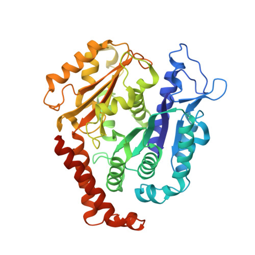
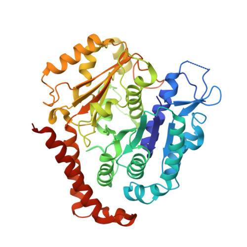
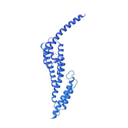
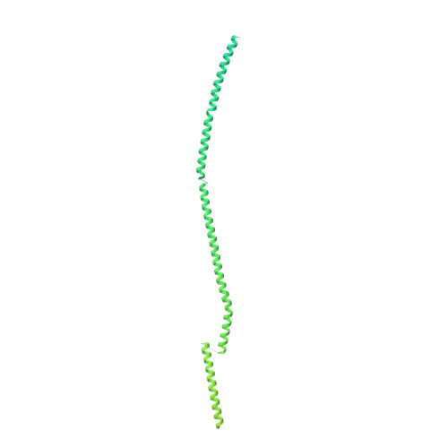
Structure of a microtubule-bound axonemal dynein.

Walton, T.Wu, H.Brown, A.

(2021) Nat Commun 12: 477-477

  • DOI: https://doi.org/10.1038/s41467-020-20735-7
  • Primary Citation of Related Structures:  
    7KZM, 7KZN, 7KZO

  • PubMed Abstract: 

    Axonemal dyneins are tethered to doublet microtubules inside cilia to drive ciliary beating, a process critical for cellular motility and extracellular fluid flow. Axonemal dyneins are evolutionarily and biochemically distinct from cytoplasmic dyneins that transport cargo, and the mechanisms regulating their localization and function are poorly understood. Here, we report a single-particle cryo-EM reconstruction of a three-headed axonemal dynein natively bound to doublet microtubules isolated from cilia. The slanted conformation of the axonemal dynein causes interaction of its motor domains with the neighboring dynein complex. Our structure shows how a heterotrimeric docking complex specifically localizes the linear array of axonemal dyneins to the doublet microtubule by directly interacting with the heavy chains. Our structural analysis establishes the arrangement of conserved heavy, intermediate and light chain subunits, and provides a framework to understand the roles of individual subunits and the interactions between dyneins during ciliary waveform generation.


  • Organizational Affiliation

    Department of Biological Chemistry and Molecular Pharmacology, Blavatnik Institute, Harvard Medical School, 240 Longwood Avenue, Boston, MA, USA.


Macromolecules
Find similar proteins by:  (by identity cutoff)  |  3D Structure
Entity ID: 1
MoleculeChains Sequence LengthOrganismDetailsImage
Tubulin beta443Chlamydomonas reinhardtiiMutation(s): 0 
UniProt
Find proteins for P04690 (Chlamydomonas reinhardtii)
Explore P04690 
Go to UniProtKB:  P04690
Entity Groups  
Sequence Clusters30% Identity50% Identity70% Identity90% Identity95% Identity100% Identity
UniProt GroupP04690
Sequence Annotations
Expand
  • Reference Sequence
Find similar proteins by:  (by identity cutoff)  |  3D Structure
Entity ID: 2
MoleculeChains Sequence LengthOrganismDetailsImage
Tubulin alpha451Chlamydomonas reinhardtiiMutation(s): 0 
UniProt
Find proteins for P09204 (Chlamydomonas reinhardtii)
Explore P09204 
Go to UniProtKB:  P09204
Entity Groups  
Sequence Clusters30% Identity50% Identity70% Identity90% Identity95% Identity100% Identity
UniProt GroupP09204
Sequence Annotations
Expand
  • Reference Sequence
Find similar proteins by:  (by identity cutoff)  |  3D Structure
Entity ID: 3
MoleculeChains Sequence LengthOrganismDetailsImage
Dynein gamma chain, flagellar outer armO [auth C]4,485Chlamydomonas reinhardtiiMutation(s): 0 
UniProt
Find proteins for Q39575 (Chlamydomonas reinhardtii)
Explore Q39575 
Go to UniProtKB:  Q39575
Entity Groups  
Sequence Clusters30% Identity50% Identity70% Identity90% Identity95% Identity100% Identity
UniProt GroupQ39575
Sequence Annotations
Expand
  • Reference Sequence
Find similar proteins by:  (by identity cutoff)  |  3D Structure
Entity ID: 4
MoleculeChains Sequence LengthOrganismDetailsImage
Outer dynein arm-docking complex subunit 1P [auth X],
R [auth X1]
749Chlamydomonas reinhardtiiMutation(s): 0 
EC: 2.7.1
UniProt
Find proteins for A8IPZ5 (Chlamydomonas reinhardtii)
Explore A8IPZ5 
Go to UniProtKB:  A8IPZ5
Entity Groups  
Sequence Clusters30% Identity50% Identity70% Identity90% Identity95% Identity100% Identity
UniProt GroupA8IPZ5
Sequence Annotations
Expand
  • Reference Sequence
Find similar proteins by:  (by identity cutoff)  |  3D Structure
Entity ID: 5
MoleculeChains Sequence LengthOrganismDetailsImage
Outer dynein arm protein 1Q [auth Y],
S [auth Y1]
552Chlamydomonas reinhardtiiMutation(s): 0 
UniProt
Find proteins for A8JF70 (Chlamydomonas reinhardtii)
Explore A8JF70 
Go to UniProtKB:  A8JF70
Entity Groups  
Sequence Clusters30% Identity50% Identity70% Identity90% Identity95% Identity100% Identity
UniProt GroupA8JF70
Sequence Annotations
Expand
  • Reference Sequence
Find similar proteins by:  (by identity cutoff)  |  3D Structure
Entity ID: 6
MoleculeChains Sequence LengthOrganismDetailsImage
Outer dynein arm-docking complex protein DC3T [auth Z]184Chlamydomonas reinhardtiiMutation(s): 0 
UniProt
Find proteins for Q7Y0H2 (Chlamydomonas reinhardtii)
Explore Q7Y0H2 
Go to UniProtKB:  Q7Y0H2
Entity Groups  
Sequence Clusters30% Identity50% Identity70% Identity90% Identity95% Identity100% Identity
UniProt GroupQ7Y0H2
Sequence Annotations
Expand
  • Reference Sequence
Small Molecules
Ligands 3 Unique
IDChains Name / Formula / InChI Key2D Diagram3D Interactions
GTP
Query on GTP

Download Ideal Coordinates CCD File 
BA [auth A5]
EA [auth A7]
HA [auth B2]
LA [auth B5]
OA [auth B7]
BA [auth A5],
EA [auth A7],
HA [auth B2],
LA [auth B5],
OA [auth B7],
U [auth A1],
Y [auth A3]
GUANOSINE-5'-TRIPHOSPHATE
C10 H16 N5 O14 P3
XKMLYUALXHKNFT-UUOKFMHZSA-N
GDP
Query on GDP

Download Ideal Coordinates CCD File 
CA [auth A5]
FA [auth A7]
GA [auth B1]
JA [auth B3]
MA [auth B5]
CA [auth A5],
FA [auth A7],
GA [auth B1],
JA [auth B3],
MA [auth B5],
PA [auth B7],
W [auth A1],
Z [auth A3]
GUANOSINE-5'-DIPHOSPHATE
C10 H15 N5 O11 P2
QGWNDRXFNXRZMB-UUOKFMHZSA-N
MG
Query on MG

Download Ideal Coordinates CCD File 
AA [auth A4]
DA [auth A6]
IA [auth B3]
KA [auth B4]
NA [auth B6]
AA [auth A4],
DA [auth A6],
IA [auth B3],
KA [auth B4],
NA [auth B6],
V [auth A1],
X [auth A2]
MAGNESIUM ION
Mg
JLVVSXFLKOJNIY-UHFFFAOYSA-N
Experimental Data & Validation

Experimental Data

  • Method: ELECTRON MICROSCOPY
  • Resolution: 3.30 Å
  • Aggregation State: FILAMENT 
  • Reconstruction Method: HELICAL 
EM Software:
TaskSoftware PackageVersion
MODEL REFINEMENTPHENIX1.18.2
RECONSTRUCTIONRELION3.1

Structure Validation

View Full Validation Report


Entry History 

Deposition Data

Revision History  (Full details and data files)

  • Version 1.0: 2021-01-20
    Type: Initial release
  • Version 1.1: 2021-02-03
    Changes: Database references
  • Version 1.2: 2024-03-06
    Changes: Data collection, Database references