7KS9 | pdb_00007ks9

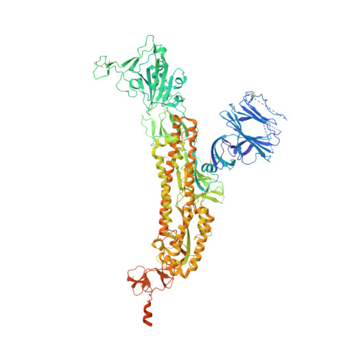
Cryo-EM structure of prefusion SARS-CoV-2 spike glycoprotein in complex with 910-30 Fab


Experimental Data Snapshot

  • Method: ELECTRON MICROSCOPY
  • Resolution: 4.75 Å
  • Aggregation State: PARTICLE 
  • Reconstruction Method: SINGLE PARTICLE 

Starting Models: experimental
View more details

wwPDB Validation 3D Report Full Report

Validation slider image for 7KS9

This is version 1.3 of the entry. See complete history. 

Literature

Paired heavy- and light-chain signatures contribute to potent SARS-CoV-2 neutralization in public antibody responses.

Banach, B.B., Cerutti, G., Fahad, A.S., Shen, C.H., Oliveira De Souza, M., Katsamba, P.S., Tsybovsky, Y., Wang, P., Nair, M.S., Huang, Y., Francino-Urdaniz, I.M., Steiner, P.J., Gutierrez-Gonzalez, M., Liu, L., Lopez Acevedo, S.N., Nazzari, A.F., Wolfe, J.R., Luo, Y., Olia, A.S., Teng, I.T., Yu, J., Zhou, T., Reddem, E.R., Bimela, J., Pan, X., Madan, B., Laflin, A.D., Nimrania, R., Yuen, K.Y., Whitehead, T.A., Ho, D.D., Kwong, P.D., Shapiro, L., DeKosky, B.J.

(2021) Cell Rep 37: 109771-109771

  • DOI: https://doi.org/10.1016/j.celrep.2021.109771
  • Primary Citation Related Structures: 
    7KS9

  • PubMed Abstract: 

    Understanding mechanisms of protective antibody recognition can inform vaccine and therapeutic strategies against SARS-CoV-2. We report a monoclonal antibody, 910-30, targeting the SARS-CoV-2 receptor-binding site for ACE2 as a member of a public antibody response encoded by IGHV3-53/IGHV3-66 genes. Sequence and structural analyses of 910-30 and related antibodies explore how class recognition features correlate with SARS-CoV-2 neutralization. Cryo-EM structures of 910-30 bound to the SARS-CoV-2 spike trimer reveal binding interactions and its ability to disassemble spike. Despite heavy-chain sequence similarity, biophysical analyses of IGHV3-53/3-66-encoded antibodies highlight the importance of native heavy:light pairings for ACE2-binding competition and SARS-CoV-2 neutralization. We develop paired heavy:light class sequence signatures and determine antibody precursor prevalence to be ∼1 in 44,000 human B cells, consistent with public antibody identification in several convalescent COVID-19 patients. These class signatures reveal genetic, structural, and functional immune features that are helpful in accelerating antibody-based medical interventions for SARS-CoV-2.


  • Organizational Affiliation: 
    • Bioengineering Graduate Program, University of Kansas, Lawrence, KS 66045, USA.

Macromolecule Content 

  • Total Structure Weight: 486.96 kDa 
  • Atom Count: 25,380 
  • Modeled Residue Count: 3,189 
  • Deposited Residue Count: 4,299 
  • Unique protein chains: 3

Macromolecules

Find similar proteins by:|  3D Structure
Entity ID: 1
MoleculeChains  Sequence LengthOrganismDetailsImage
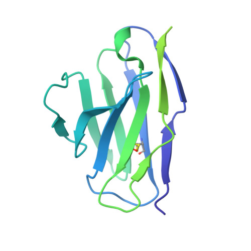
910-30 Fab light chainA [auth L]214Homo sapiensMutation(s): 0 
Entity Groups
Sequence Clusters30% Identity50% Identity70% Identity90% Identity95% Identity100% Identity
Sequence Annotations
Expand
Reference Sequence
Find similar proteins by:|  3D Structure
Entity ID: 2
MoleculeChains  Sequence LengthOrganismDetailsImage
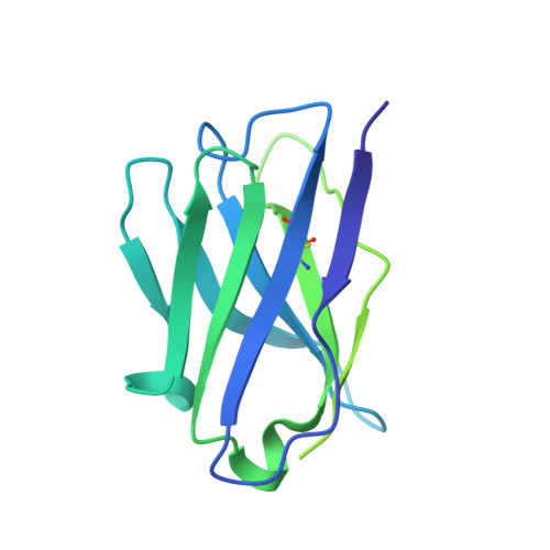
910-30 Fab heavy chainB [auth H]221Homo sapiensMutation(s): 0 
Entity Groups
Sequence Clusters30% Identity50% Identity70% Identity90% Identity95% Identity100% Identity
Sequence Annotations
Expand
Reference Sequence
Find similar proteins by:|  3D Structure
Entity ID: 3
MoleculeChains  Sequence LengthOrganismDetailsImage
Spike glycoproteinC [auth A],
D [auth B],
E [auth C]
1,288Severe acute respiratory syndrome coronavirus 2Mutation(s): 5 
Gene Names: S, 2
UniProt
Find proteins for P0DTC2 (Severe acute respiratory syndrome coronavirus 2)
Explore P0DTC2 
Go to UniProtKB:  P0DTC2
Entity Groups
Sequence Clusters30% Identity50% Identity70% Identity90% Identity95% Identity100% Identity
UniProt GroupP0DTC2
Glycosylation
Glycosylation Sites: 15Go to GlyGen: P0DTC2-1
Sequence Annotations
Expand
Reference Sequence

Oligosaccharides

Help  
Entity ID: 4
MoleculeChains Length2D Diagram GlycosylationD Interactions
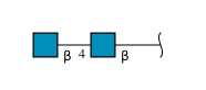
2-acetamido-2-deoxy-beta-D-glucopyranose-(1-4)-2-acetamido-2-deoxy-beta-D-glucopyranose
F [auth D],
G [auth E],
H [auth F],
I [auth G],
J [auth I],
F [auth D],
G [auth E],
H [auth F],
I [auth G],
J [auth I],
K [auth J],
L [auth K],
M,
N,
O,
P,
Q
2N-Glycosylation
Glycosylation Resources
GlyTouCan: G42666HT
GlyCosmos: G42666HT
GlyGen: G42666HT

Small Molecules

Ligands 1 Unique
IDChains Name / Formula / InChI Key2D Diagram3D Interactions
NAG
(Subject of Investigation/LOI)

Query on NAG


Download:Ideal Coordinates CCD File
AA [auth A]
BA [auth B]
CA [auth B]
DA [auth B]
EA [auth B]
AA [auth A],
BA [auth B],
CA [auth B],
DA [auth B],
EA [auth B],
FA [auth B],
GA [auth B],
HA [auth B],
IA [auth B],
JA [auth B],
KA [auth B],
LA [auth B],
MA [auth B],
NA [auth B],
OA [auth C],
PA [auth C],
QA [auth C],
R [auth A],
RA [auth C],
S [auth A],
SA [auth C],
T [auth A],
TA [auth C],
U [auth A],
UA [auth C],
V [auth A],
VA [auth C],
W [auth A],
WA [auth C],
X [auth A],
XA [auth C],
Y [auth A],
YA [auth C],
Z [auth A],
ZA [auth C]
2-acetamido-2-deoxy-beta-D-glucopyranose
C8 H15 N O6
OVRNDRQMDRJTHS-FMDGEEDCSA-N

Experimental Data & Validation

Experimental Data

  • Method: ELECTRON MICROSCOPY
  • Resolution: 4.75 Å
  • Aggregation State: PARTICLE 
  • Reconstruction Method: SINGLE PARTICLE 
EM Software:
TaskSoftware PackageVersion
RECONSTRUCTIONcryoSPARC

Structure Validation

View Full Validation Report


Entry History 

& Funding Information

Deposition Data


Funding OrganizationLocationGrant Number
Other privateChinaJack Ma Foundation

Revision History  (Full details and data files)

  • Version 1.0: 2021-02-10
    Type: Initial release
  • Version 1.1: 2021-02-17
    Changes: Database references
  • Version 1.2: 2022-11-09
    Changes: Database references
  • Version 1.3: 2024-11-13
    Changes: Data collection, Refinement description, Structure summary