7KBT | pdb_00007kbt

Factor VIII in complex with the anti-C2 domain antibody, G99


Experimental Data Snapshot

  • Method: X-RAY DIFFRACTION
  • Resolution: 4.15 Å
  • R-Value Free: 
    0.338 (Depositor), 0.338 (DCC) 
  • R-Value Work: 
    0.300 (Depositor), 0.303 (DCC) 
  • R-Value Observed: 
    0.302 (Depositor) 

Starting Model: experimental
View more details

wwPDB Validation 3D Report Full Report

Validation slider image for 7KBT

Ligand Structure Quality Assessment 


This is version 1.3 of the entry. See complete history

Literature

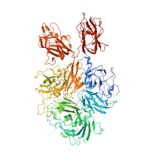
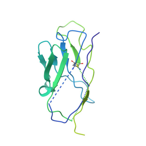
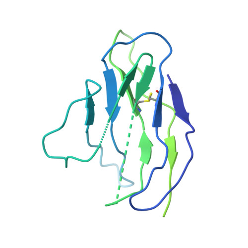
Structure of Blood Coagulation Factor VIII in Complex With an Anti-C2 Domain Non-Classical, Pathogenic Antibody Inhibitor

Ronayne, E.K.Peters, S.C.Gish, J.S.Wilson, C.Spencer, H.T.Doering, C.B.Lollar, P.Spiegel, P.C.Childers, K.C.

(2021) Front Immunol 12: 2260

Macromolecule Content 

  • Total Structure Weight: 217.12 kDa 
  • Atom Count: 11,366 
  • Modeled Residue Count: 1,444 
  • Deposited Residue Count: 1,905 
  • Unique protein chains: 3

Macromolecules

Find similar proteins by:|  3D Structure
Entity ID: 1
MoleculeChains  Sequence LengthOrganismDetailsImage
Coagulation factor VIII,Coagulation factor VIII,Coagulation factor VIII,Coagulation factor VIII,Coagulation factor VIII1,467Sus scrofaHomo sapiensMutation(s): 0 
Gene Names: F8CF8F8C
UniProt & NIH Common Fund Data Resources
Find proteins for P00451 (Homo sapiens)
Explore P00451 
Go to UniProtKB:  P00451
PHAROS:  P00451
GTEx:  ENSG00000185010 
Find proteins for P12263 (Sus scrofa)
Explore P12263 
Go to UniProtKB:  P12263
Entity Groups
Sequence Clusters30% Identity50% Identity70% Identity90% Identity95% Identity100% Identity
UniProt GroupsP12263P00451
Glycosylation
Glycosylation Sites: 3Go to GlyGen: P00451-1
Sequence Annotations
Expand
Reference Sequence
Find similar proteins by:|  3D Structure
Entity ID: 2
MoleculeChains  Sequence LengthOrganismDetailsImage
G99 heavy chainB [auth E]224Mus musculusMutation(s): 0 
Find similar proteins by:|  3D Structure
Entity ID: 3
MoleculeChains  Sequence LengthOrganismDetailsImage
G99 light chainC [auth F]214Mus musculusMutation(s): 0 

Oligosaccharides

Help  
Entity ID: 4
MoleculeChains Length2D Diagram GlycosylationD Interactions
2-acetamido-2-deoxy-beta-D-glucopyranose-(1-4)-2-acetamido-2-deoxy-beta-D-glucopyranoseD [auth G],
E [auth B]
2N-Glycosylation
Glycosylation Resources
GlyTouCan: G42666HT
GlyCosmos: G42666HT
GlyGen: G42666HT

Experimental Data & Validation

Experimental Data

  • Method: X-RAY DIFFRACTION
  • Resolution: 4.15 Å
  • R-Value Free:  0.338 (Depositor), 0.338 (DCC) 
  • R-Value Work:  0.300 (Depositor), 0.303 (DCC) 
  • R-Value Observed: 0.302 (Depositor) 
Space Group: P 41 21 2
Unit Cell:
Length ( Å )Angle ( ˚ )
a = 132.243α = 90
b = 132.243β = 90
c = 380.029γ = 90
Software Package:
Software NamePurpose
PHENIXrefinement
Adxvdata reduction
XDSdata reduction
PHASERphasing
Aimlessdata scaling
Cootmodel building

Structure Validation

View Full Validation Report


Ligand Structure Quality Assessment 


Entry History 

& Funding Information

Deposition Data


Funding OrganizationLocationGrant Number
National Science Foundation (NSF, United States)United StatesMRI 1429164
National Institutes of Health/National Heart, Lung, and Blood Institute (NIH/NHLBI)United StatesR15HL103518
National Institutes of Health/National Heart, Lung, and Blood Institute (NIH/NHLBI)United StatesU54HL141981
National Institutes of Health/National Heart, Lung, and Blood Institute (NIH/NHLBI)United StatesR44HL117511
National Institutes of Health/National Heart, Lung, and Blood Institute (NIH/NHLBI)United StatesR44HL110448
National Institutes of Health/National Heart, Lung, and Blood Institute (NIH/NHLBI)United StatesU54HL112309

Revision History  (Full details and data files)

  • Version 1.0: 2020-11-18
    Type: Initial release
  • Version 1.1: 2021-06-16
    Changes: Database references
  • Version 1.2: 2023-10-18
    Changes: Data collection, Database references, Refinement description
  • Version 1.3: 2024-10-23
    Changes: Structure summary