


Crystal Structure of PH0140 from Pyrococcus horikosii OT3
Richard, M., Ahmad, M., Pal, R.K., Biswal, B.K., Jeyakanthan, J.To be published.
Experimental Data Snapshot
Starting Model: experimental
View more details
Entity ID: 1 | |||||
|---|---|---|---|---|---|
| Molecule | Chains | Sequence Length | Organism | Details | Image |
| Transcriptional regulatory protein | 164 | Pyrococcus horikoshii OT3 | Mutation(s): 0 Gene Names: PH0140 | 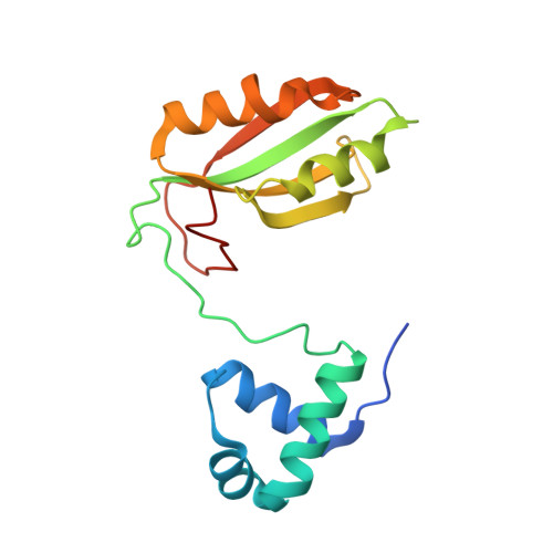 | |
UniProt | |||||
Entity Groups | |||||
| Sequence Clusters | 30% Identity50% Identity70% Identity90% Identity95% Identity100% Identity | ||||
| UniProt Group | O57880 | ||||
Sequence AnnotationsExpand | |||||
Reference Sequence | |||||
| Ligands 2 Unique | |||||
|---|---|---|---|---|---|
| ID | Chains | Name / Formula / InChI Key | 2D Diagram | 3D Interactions | |
| ILE (Subject of Investigation/LOI) Download:Ideal Coordinates CCD File | B [auth A] | ISOLEUCINE C6 H13 N O2 AGPKZVBTJJNPAG-WHFBIAKZSA-N |  | ||
| EDO Download:Ideal Coordinates CCD File | C [auth A], D [auth A] | 1,2-ETHANEDIOL C2 H6 O2 LYCAIKOWRPUZTN-UHFFFAOYSA-N |  | ||
| Length ( Å ) | Angle ( ˚ ) |
|---|---|
| a = 102.79 | α = 90 |
| b = 102.79 | β = 90 |
| c = 69.183 | γ = 90 |
| Software Name | Purpose |
|---|---|
| REFMAC | refinement |
| HKL-2000 | data reduction |
| HKL-2000 | data scaling |
| PHASER | phasing |


