7E50

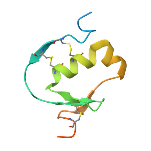
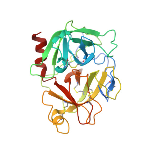
Crystal structure of human microplasmin in complex with kazal-type inhibitor AaTI


Experimental Data Snapshot

  • Method: X-RAY DIFFRACTION
  • Resolution: 1.95 Å
  • R-Value Free: 0.275 
  • R-Value Work: 0.218 
  • R-Value Observed: 0.222 

wwPDB Validation   3D Report Full Report


This is version 1.2 of the entry. See complete history


Literature

Crystal structure of Aedes aegypti trypsin inhibitor in complex with mu-plasmin reveals role for scaffold stability in Kazal-type serine protease inhibitor.

Walvekar, V.A.Ramesh, K.Jobichen, C.Kannan, M.Sivaraman, J.Kini, R.M.Mok, Y.K.

(2022) Protein Sci 31: 470-484

  • DOI: https://doi.org/10.1002/pro.4245
  • Primary Citation of Related Structures:  
    7E50

  • PubMed Abstract: 

    Kazal-type protease inhibitor specificity is believed to be determined by sequence of the reactive-site loop that make most, if not all, contacts with the serine protease. Here, we determined the complex crystal structure of Aedes aegypti trypsin inhibitor (AaTI) with μ-plasmin, and compared its reactivities with other Kazal-type inhibitors, infestin-1 and infestin-4. We show that the shortened 99-loop of plasmin creates an S2 pocket, which is filled by phenylalanine at the P2 position of the reactive-site loop of infestin-4. In contrast, AaTI and infestin-1 retain a proline at P2, rendering the S2 pocket unfilled, which leads to lower plasmin inhibitions. Furthermore, the protein scaffold of AaTI is unstable, due to an elongated Cys-V to Cys-VI region leading to a less compact hydrophobic core. Chimeric study shows that the stability of the scaffold can be modified by swapping of this Cys-V to Cys-VI region between AaTI and infestin-4. The scaffold instability causes steric clashing of the bulky P2 residue, leading to significantly reduced inhibition of plasmin by AaTI or infestin-4 chimera. Our findings suggest that surface loops of protease and scaffold stability of Kazal-type inhibitor are both necessary for specific protease inhibition, in addition to reactive site loop sequence. PDB ID code: 7E50.


  • Organizational Affiliation

    Department of Biological Sciences, National University of Singapore, Singapore.


Macromolecules
Find similar proteins by:  (by identity cutoff)  |  3D Structure
Entity ID: 1
MoleculeChains Sequence LengthOrganismDetailsImage
AAEL006007-PA79Aedes aegyptiMutation(s): 0 
Gene Names: AAEL006007
UniProt
Find proteins for A0A1S4FCI9 (Aedes aegypti)
Explore A0A1S4FCI9 
Go to UniProtKB:  A0A1S4FCI9
Entity Groups  
Sequence Clusters30% Identity50% Identity70% Identity90% Identity95% Identity100% Identity
UniProt GroupA0A1S4FCI9
Sequence Annotations
Expand
  • Reference Sequence
Find similar proteins by:  (by identity cutoff)  |  3D Structure
Entity ID: 2
MoleculeChains Sequence LengthOrganismDetailsImage
Plasminogen255Homo sapiensMutation(s): 0 
Gene Names: PLG
EC: 3.4.21.7
UniProt & NIH Common Fund Data Resources
Find proteins for P00747 (Homo sapiens)
Explore P00747 
Go to UniProtKB:  P00747
PHAROS:  P00747
GTEx:  ENSG00000122194 
Entity Groups  
Sequence Clusters30% Identity50% Identity70% Identity90% Identity95% Identity100% Identity
UniProt GroupP00747
Sequence Annotations
Expand
  • Reference Sequence
Experimental Data & Validation

Experimental Data

  • Method: X-RAY DIFFRACTION
  • Resolution: 1.95 Å
  • R-Value Free: 0.275 
  • R-Value Work: 0.218 
  • R-Value Observed: 0.222 
  • Space Group: P 21 21 2
Unit Cell:
Length ( Å )Angle ( ˚ )
a = 72.895α = 90
b = 86.044β = 90
c = 50.315γ = 90
Software Package:
Software NamePurpose
PHENIXrefinement
PDB_EXTRACTdata extraction
HKL-2000data reduction
HKL-2000data scaling
PHASERphasing

Structure Validation

View Full Validation Report


Entry History 

Deposition Data

Revision History  (Full details and data files)

  • Version 1.0: 2022-02-16
    Type: Initial release
  • Version 1.1: 2023-08-16
    Changes: Data collection, Database references
  • Version 1.2: 2023-11-29
    Changes: Refinement description