7E50

Crystal structure of human microplasmin in complex with kazal-type inhibitor AaTI


Experimental Data Snapshot

  • Method: X-RAY DIFFRACTION
  • Resolution: 1.95 Å
  • R-Value Free: 0.275 
  • R-Value Work: 0.218 
  • R-Value Observed: 0.222 

Starting Model: experimental
View more details

wwPDB Validation   3D Report Full Report


This is version 1.2 of the entry. See complete history


Literature

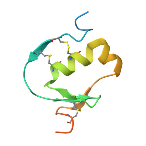
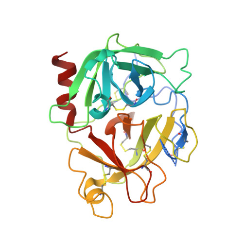
Crystal structure of Aedes aegypti trypsin inhibitor in complex with mu-plasmin reveals role for scaffold stability in Kazal-type serine protease inhibitor.

Walvekar, V.A.Ramesh, K.Jobichen, C.Kannan, M.Sivaraman, J.Kini, R.M.Mok, Y.K.

(2022) Protein Sci 31: 470-484

  • DOI: https://doi.org/10.1002/pro.4245
  • Primary Citation of Related Structures:  
    7E50

  • PubMed Abstract: 

    Kazal-type protease inhibitor specificity is believed to be determined by sequence of the reactive-site loop that make most, if not all, contacts with the serine protease. Here, we determined the complex crystal structure of Aedes aegypti trypsin inhibitor (AaTI) with μ-plasmin, and compared its reactivities with other Kazal-type inhibitors, infestin-1 and infestin-4. We show that the shortened 99-loop of plasmin creates an S2 pocket, which is filled by phenylalanine at the P2 position of the reactive-site loop of infestin-4. In contrast, AaTI and infestin-1 retain a proline at P2, rendering the S2 pocket unfilled, which leads to lower plasmin inhibitions. Furthermore, the protein scaffold of AaTI is unstable, due to an elongated Cys-V to Cys-VI region leading to a less compact hydrophobic core. Chimeric study shows that the stability of the scaffold can be modified by swapping of this Cys-V to Cys-VI region between AaTI and infestin-4. The scaffold instability causes steric clashing of the bulky P2 residue, leading to significantly reduced inhibition of plasmin by AaTI or infestin-4 chimera. Our findings suggest that surface loops of protease and scaffold stability of Kazal-type inhibitor are both necessary for specific protease inhibition, in addition to reactive site loop sequence. PDB ID code: 7E50.


  • Organizational Affiliation

    Department of Biological Sciences, National University of Singapore, Singapore.


Macromolecules
Find similar proteins by:  (by identity cutoff)  |  3D Structure
Entity ID: 1
MoleculeChains Sequence LengthOrganismDetailsImage
AAEL006007-PA79Aedes aegyptiMutation(s): 0 
Gene Names: AAEL006007
UniProt
Find proteins for A0A1S4FCI9 (Aedes aegypti)
Explore A0A1S4FCI9 
Go to UniProtKB:  A0A1S4FCI9
Entity Groups  
Sequence Clusters30% Identity50% Identity70% Identity90% Identity95% Identity100% Identity
UniProt GroupA0A1S4FCI9
Sequence Annotations
Expand
  • Reference Sequence
Find similar proteins by:  (by identity cutoff)  |  3D Structure
Entity ID: 2
MoleculeChains Sequence LengthOrganismDetailsImage
Plasminogen255Homo sapiensMutation(s): 0 
Gene Names: PLG
EC: 3.4.21.7
UniProt & NIH Common Fund Data Resources
Find proteins for P00747 (Homo sapiens)
Explore P00747 
Go to UniProtKB:  P00747
PHAROS:  P00747
GTEx:  ENSG00000122194 
Entity Groups  
Sequence Clusters30% Identity50% Identity70% Identity90% Identity95% Identity100% Identity
UniProt GroupP00747
Sequence Annotations
Expand
  • Reference Sequence
Experimental Data & Validation

Experimental Data

  • Method: X-RAY DIFFRACTION
  • Resolution: 1.95 Å
  • R-Value Free: 0.275 
  • R-Value Work: 0.218 
  • R-Value Observed: 0.222 
  • Space Group: P 21 21 2
Unit Cell:
Length ( Å )Angle ( ˚ )
a = 72.895α = 90
b = 86.044β = 90
c = 50.315γ = 90
Software Package:
Software NamePurpose
PHENIXrefinement
PDB_EXTRACTdata extraction
HKL-2000data reduction
HKL-2000data scaling
PHASERphasing

Structure Validation

View Full Validation Report


Entry History 

Deposition Data

Revision History  (Full details and data files)

  • Version 1.0: 2022-02-16
    Type: Initial release
  • Version 1.1: 2023-08-16
    Changes: Data collection, Database references
  • Version 1.2: 2023-11-29
    Changes: Refinement description