Asymmetric total synthesis of polycyclic xanthenes and discovery of a WalK activator active against MRSA.
Cheng, M.J., Wu, Y.Y., Zeng, H., Zhang, T.H., Hu, Y.X., Liu, S.Y., Cui, R.Q., Hu, C.X., Zou, Q.M., Li, C.C., Ye, W.C., Huang, W., Wang, L.(2024) Nat Commun 15: 5879-5879
- PubMed: 38997253 Search on PubMedSearch on PubMed Central
- DOI: https://doi.org/10.1038/s41467-024-49629-8
- Primary Citation Related Structures:
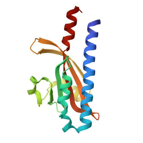
7DUD - PubMed Abstract:
The development of new antibiotics continues to pose challenges, particularly considering the growing threat of multidrug-resistant Staphylococcus aureus. Structurally diverse natural products provide a promising source of antibiotics. Herein, we outline a concise approach for the collective asymmetric total synthesis of polycyclic xanthene myrtucommulone D and five related congeners. The strategy involves rapid assembly of the challenging benzopyrano[2,3-a]xanthene core, highly diastereoselective establishment of three contiguous stereocenters through a retro-hemiketalization/double Michael cascade reaction, and a Mitsunobu-mediated chiral resolution approach with high optical purity and broad substrate scope. Quantum mechanical calculations provide insight into stereoselective construction mechanism of the three contiguous stereocenters. Additionally, this work leads to the discovery of an antibacterial agent against both drug-sensitive and drug-resistant S. aureus. This compound operates through a unique mechanism that promotes bacterial autolysis by activating the two-component sensory histidine kinase WalK. Our research holds potential for future antibacterial drug development.
- State Key Laboratory of Bioactive Molecules and Druggability Assessment, Jinan University, Guangzhou, 510632, P. R. China.
Organizational Affiliation: