7DER

Lysozyme alone in H2O

  • Classification: HYDROLASE
  • Organism(s): Gallus gallus
  • Mutation(s): No 

  • Deposited: 2020-11-04 Released: 2021-03-10 
  • Deposition Author(s): Tanaka, I., Chatake, T.
  • Funding Organization(s): Japan Society for the Promotion of Science (JSPS)

Experimental Data Snapshot

  • Method: X-RAY DIFFRACTION
  • Resolution: 1.03 Å
  • R-Value Free: 0.173 
  • R-Value Work: 0.149 
  • R-Value Observed: 0.149 

wwPDB Validation   3D Report Full Report


This is version 1.1 of the entry. See complete history


Literature

Recent structural insights into the mechanism of lysozyme hydrolysis.

Tanaka, I.Nishinomiya, R.Goto, R.Shimazaki, S.Chatake, T.

(2021) Acta Crystallogr D Struct Biol 77: 288-292

  • DOI: https://doi.org/10.1107/S2059798321000346
  • Primary Citation of Related Structures:  
    7BR5, 7DEQ, 7DER

  • PubMed Abstract: 

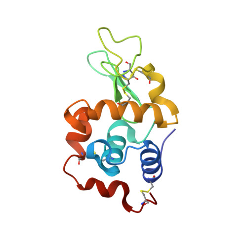
    Lysozyme hydrolyzes the glycosidic bonds between N-acetylmuramic acid and N-acetylglucosamine in peptidoglycans located in the bacterial cell wall. The mechanism of the hydrolysis reaction of lysozyme was first studied more than 50 years ago; however, it has not yet been fully elucidated and various mechanisms are still being investigated. One reaction system that has commonly been proposed is that the lysozyme intermediate undergoes covalent ligand binding during hydrolysis. However, these findings resulted from experiments performed under laboratory conditions using fluorine-based ligands, which facilitate the formation of covalent bonds between the ligands and the catalytic side chain of lysozyme. More recently, high-resolution X-ray structural analysis was used to study the complex of lysozyme with an N-acetylglucosamine tetramer. As a result, the carboxyl group of Asp52 was found to form a relatively strong hydrogen-bond network and had difficulty binding covalently to C1 of the carbohydrate ring. To confirm this hydrogen-bond network, neutron test measurements were successfully performed to a resolution of better than 1.9 Å.


  • Organizational Affiliation

    Graduate School of Science and Engineering, Ibaraki University, Hitachi, Ibaraki 316-8511, Japan.


Macromolecules
Find similar proteins by:  (by identity cutoff)  |  3D Structure
Entity ID: 1
MoleculeChains Sequence LengthOrganismDetailsImage
Lysozyme C129Gallus gallusMutation(s): 0 
EC: 3.2.1.17
UniProt
Find proteins for P00698 (Gallus gallus)
Explore P00698 
Go to UniProtKB:  P00698
Entity Groups  
Sequence Clusters30% Identity50% Identity70% Identity90% Identity95% Identity100% Identity
UniProt GroupP00698
Sequence Annotations
Expand
  • Reference Sequence
Experimental Data & Validation

Experimental Data

  • Method: X-RAY DIFFRACTION
  • Resolution: 1.03 Å
  • R-Value Free: 0.173 
  • R-Value Work: 0.149 
  • R-Value Observed: 0.149 
  • Space Group: P 43 21 2
Unit Cell:
Length ( Å )Angle ( ˚ )
a = 78.777α = 90
b = 78.777β = 90
c = 37.093γ = 90
Software Package:
Software NamePurpose
PHENIXrefinement
XDSdata reduction
Aimlessdata scaling
Cootmodel building

Structure Validation

View Full Validation Report


Entry History & Funding Information

Deposition Data


Funding OrganizationLocationGrant Number
Japan Society for the Promotion of Science (JSPS)Japan20H05436

Revision History  (Full details and data files)

  • Version 1.0: 2021-03-10
    Type: Initial release
  • Version 1.1: 2023-11-29
    Changes: Data collection, Database references, Refinement description