7CP1

Crystal structure of isocitrate lyase in complex with succinate and itaconate


Experimental Data Snapshot

  • Method: X-RAY DIFFRACTION
  • Resolution: 2.58 Å
  • R-Value Free: 0.194 
  • R-Value Work: 0.154 
  • R-Value Observed: 0.156 

wwPDB Validation   3D Report Full Report


Ligand Structure Quality Assessment 


This is version 1.1 of the entry. See complete history


Literature

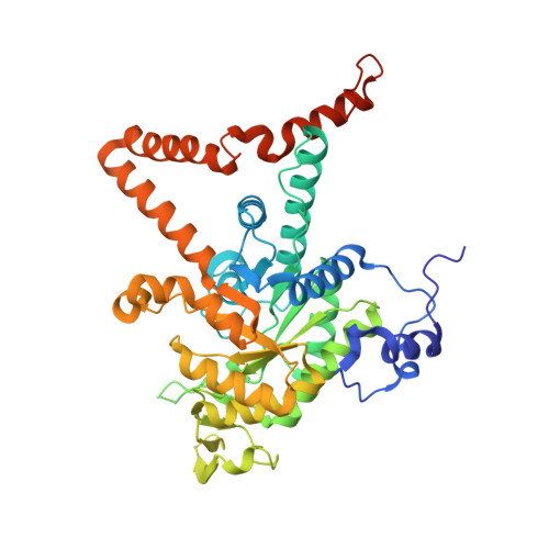
Heterogeneous multimeric structure of isocitrate lyase in complex with succinate and itaconate provides novel insights into its inhibitory mechanism.

Kwon, S.Chun, H.L.Ha, H.J.Lee, S.Y.Park, H.H.

(2021) PLoS One 16: e0251067-e0251067

  • DOI: https://doi.org/10.1371/journal.pone.0251067
  • Primary Citation of Related Structures:  
    7CP1

  • PubMed Abstract: 

    During the glyoxylate cycle, isocitrate lyases (ICLs) catalyze the lysis of isocitrate to glyoxylate and succinate. Itaconate has been reported to inhibit an ICL from Mycobacterium tuberculosis (tbICL). To elucidate the molecular mechanism of ICL inhibition, we determined the crystal structure of tbICL in complex with itaconate. Unexpectedly, succinate and itaconate were found to bind to the respective active sites in the dimeric form of tbICL. Our structure revealed the active site architecture as an open form, although the substrate and inhibitor were bound to the active sites. Our findings provide novel insights into the conformation of tbICL upon its binding to a substrate or inhibitor, along with molecular details of the inhibitory mechanism of itaconate.


  • Organizational Affiliation

    Department of Biotechnology, Konkuk University, Chungju, Chungbuk, Republic of Korea.


Macromolecules
Find similar proteins by:  (by identity cutoff)  |  3D Structure
Entity ID: 1
MoleculeChains Sequence LengthOrganismDetailsImage
Isocitrate lyase
A, B
436Mycobacterium tuberculosis H37RvMutation(s): 0 
Gene Names: iclRv0467MTV038.11
EC: 4.1.3.1
UniProt
Find proteins for P9WKK7 (Mycobacterium tuberculosis (strain ATCC 25618 / H37Rv))
Explore P9WKK7 
Go to UniProtKB:  P9WKK7
Entity Groups  
Sequence Clusters30% Identity50% Identity70% Identity90% Identity95% Identity100% Identity
UniProt GroupP9WKK7
Sequence Annotations
Expand
  • Reference Sequence
Experimental Data & Validation

Experimental Data

  • Method: X-RAY DIFFRACTION
  • Resolution: 2.58 Å
  • R-Value Free: 0.194 
  • R-Value Work: 0.154 
  • R-Value Observed: 0.156 
  • Space Group: P 65 2 2
Unit Cell:
Length ( Å )Angle ( ˚ )
a = 131.32α = 90
b = 131.32β = 90
c = 284.882γ = 120
Software Package:
Software NamePurpose
PHENIXrefinement
HKL-2000data scaling
PDB_EXTRACTdata extraction
HKL-2000data reduction
PHENIXphasing

Structure Validation

View Full Validation Report


Ligand Structure Quality Assessment 


Entry History & Funding Information

Deposition Data


Funding OrganizationLocationGrant Number
National Research Foundation (NRF, Korea)2017M3A9D8062960
National Research Foundation (NRF, Korea)Korea, Republic Of2018R1A4A1023822

Revision History  (Full details and data files)

  • Version 1.0: 2021-05-19
    Type: Initial release
  • Version 1.1: 2023-11-29
    Changes: Data collection, Database references, Refinement description