7CGW

Complex structure of PD-1 and tislelizumab Fab


Experimental Data Snapshot

  • Method: X-RAY DIFFRACTION
  • Resolution: 3.20 Å
  • R-Value Free: 0.254 
  • R-Value Work: 0.218 
  • R-Value Observed: 0.220 

wwPDB Validation   3D Report Full Report


Ligand Structure Quality Assessment 


This is version 1.1 of the entry. See complete history


Literature

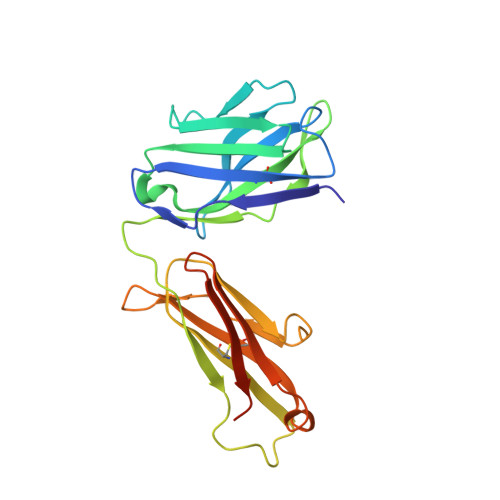
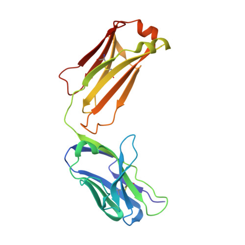
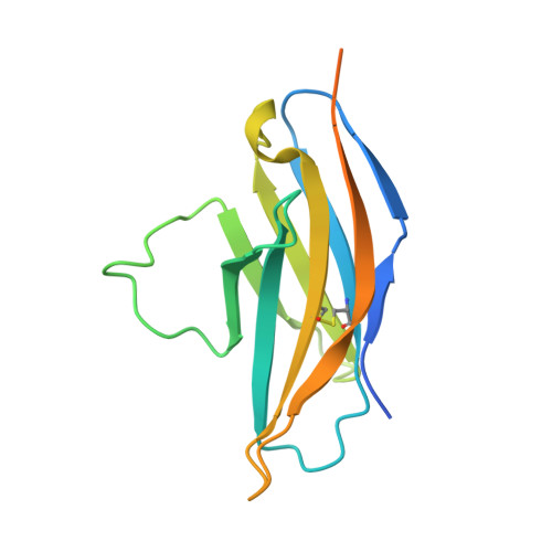
Tislelizumab uniquely binds to the CC' loop of PD-1 with slow-dissociated rate and complete PD-L1 blockage.

Hong, Y.Feng, Y.Sun, H.Zhang, B.Wu, H.Zhu, Q.Li, Y.Zhang, T.Zhang, Y.Cui, X.Li, Z.Song, X.Li, K.Liu, M.Liu, Y.

(2021) FEBS Open Bio 11: 782-792

  • DOI: https://doi.org/10.1002/2211-5463.13102
  • Primary Citation of Related Structures:  
    7CGW

  • PubMed Abstract: 

    Programmed cell death protein 1 (PD-1), an immune checkpoint receptor expressed by activated T, B, and NK cells, is a well-known target for cancer immunotherapy. Tislelizumab (BGB-A317) is an anti-PD-1 antibody that has recently been approved for treatment of Hodgkin's lymphoma and urothelial carcinoma. Here, we show that tislelizumab displayed remarkable antitumor efficacy in a B16F10/GM-CSF mouse model. Structural biology and Surface plasmon resonance (SPR) analyses revealed unique epitopes of tislelizumab, and demonstrated that the CC' loop of PD-1, a region considered to be essential for binding to PD-1 ligand 1 (PD-L1) but not reported as targeted by other therapeutic antibodies, significantly contributes to the binding of tislelizumab. The binding surface of tislelizumab on PD-1 overlaps largely with that of the PD-L1. SPR analysis revealed the extremely slow dissociation rate of tislelizumab from PD-1. Both structural and functional analyses align with the observed ability of tislelizumab to completely block PD-1/PD-L1 interaction, broadening our understanding of the mechanism of action of anti-PD-1 antibodies.


  • Organizational Affiliation

    BeiGene Global Research, BeiGene (Beijing) Co., Ltd., China.


Macromolecules
Find similar proteins by:  (by identity cutoff)  |  3D Structure
Entity ID: 1
MoleculeChains Sequence LengthOrganismDetailsImage
Heavy chain of tislelizumab FabA,
D [auth H]
228Homo sapiensMutation(s): 0 
Entity Groups  
Sequence Clusters30% Identity50% Identity70% Identity90% Identity95% Identity100% Identity
Sequence Annotations
Expand
  • Reference Sequence
Find similar proteins by:  (by identity cutoff)  |  3D Structure
Entity ID: 2
MoleculeChains Sequence LengthOrganismDetailsImage
Light chain of tislelizumab FabB,
E [auth L]
214Homo sapiensMutation(s): 0 
Entity Groups  
Sequence Clusters30% Identity50% Identity70% Identity90% Identity95% Identity100% Identity
Sequence Annotations
Expand
  • Reference Sequence
Find similar proteins by:  (by identity cutoff)  |  3D Structure
Entity ID: 3
MoleculeChains Sequence LengthOrganismDetailsImage
Programmed cell death protein 1C,
F [auth P]
151Homo sapiensMutation(s): 0 
Gene Names: PDCD1PD1
UniProt & NIH Common Fund Data Resources
Find proteins for Q15116 (Homo sapiens)
Explore Q15116 
Go to UniProtKB:  Q15116
PHAROS:  Q15116
GTEx:  ENSG00000188389 
Entity Groups  
Sequence Clusters30% Identity50% Identity70% Identity90% Identity95% Identity100% Identity
UniProt GroupQ15116
Sequence Annotations
Expand
  • Reference Sequence
Experimental Data & Validation

Experimental Data

  • Method: X-RAY DIFFRACTION
  • Resolution: 3.20 Å
  • R-Value Free: 0.254 
  • R-Value Work: 0.218 
  • R-Value Observed: 0.220 
  • Space Group: C 2 2 21
Unit Cell:
Length ( Å )Angle ( ˚ )
a = 142.638α = 90
b = 145.572β = 90
c = 175.549γ = 90
Software Package:
Software NamePurpose
PHENIXrefinement
XDSdata reduction
Aimlessdata scaling
PHENIXphasing

Structure Validation

View Full Validation Report


Ligand Structure Quality Assessment 


Entry History 

Deposition Data

Revision History  (Full details and data files)

  • Version 1.0: 2021-04-07
    Type: Initial release
  • Version 1.1: 2024-04-03
    Changes: Data collection, Database references, Refinement description