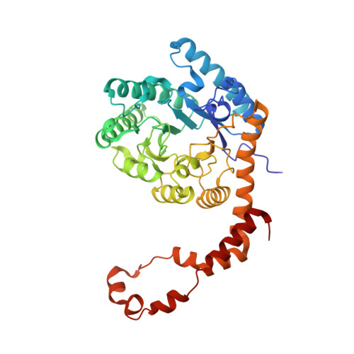
Nucleation of protein mesocrystals via oriented attachment.
Van Driessche, A.E.S., Van Gerven, N., Joosten, R.R.M., Ling, W.L., Bacia, M., Sommerdijk, N., Sleutel, M.(2021) Nat Commun 12: 3902-3902
- PubMed: 34162863 Search on PubMedSearch on PubMed Central
- DOI: https://doi.org/10.1038/s41467-021-24171-z
- Primary Citation Related Structures:
7BJZ - PubMed Abstract:
Self-assembly of proteins holds great promise for the bottom-up design and production of synthetic biomaterials. In conventional approaches, designer proteins are pre-programmed with specific recognition sites that drive the association process towards a desired organized state. Although proven effective, this approach poses restrictions on the complexity and material properties of the end-state. An alternative, hierarchical approach that has found wide adoption for inorganic systems, relies on the production of crystalline nanoparticles that become the building blocks of a next-level assembly process driven by oriented attachment (OA). As it stands, OA has not yet been observed for protein systems. Here we employ cryo-transmission electron microscopy (cryoEM) in the high nucleation rate limit of protein crystals and map the self-assembly route at molecular resolution. We observe the initial formation of facetted nanocrystals that merge lattices by means of OA alignment well before contact is made, satisfying non-trivial symmetry rules in the process. As these nanocrystalline assemblies grow larger we witness imperfect docking events leading to oriented aggregation into mesocrystalline assemblies. These observations highlight the underappreciated role of the interaction between crystalline nuclei, and the impact of OA on the crystallization process of proteins.
- Univ. Grenoble Alpes, CNRS, ISTerre, Grenoble, France.
Organizational Affiliation: