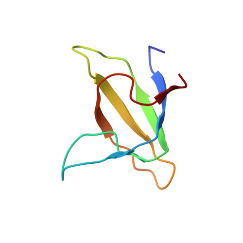
Solution NMR Structure of the SH3 Domain of Human Caskin1 Validates the Lack of a Typical Peptide Binding Groove and Supports a Role in Lipid Mediator Binding.
Toke, O., Koprivanacz, K., Radnai, L., Mero, B., Juhasz, T., Liliom, K., Buday, L.(2021) Cells 10
- PubMed: 33467043 Search on PubMedSearch on PubMed Central
- DOI: https://doi.org/10.3390/cells10010173
- Primary Citation Related Structures:
7ATY - PubMed Abstract:
SH3 domains constitute an important class of protein modules involved in a variety of cellular functions. They participate in protein-protein interactions via their canonical ligand binding interfaces composed of several evolutionarily conserved aromatic residues forming binding grooves for typical (PxxP) and atypical (PxxxPR, RxxK, RKxxY) binding motifs. The calcium/calmodulin-dependent serine protein kinase (CASK)-interacting protein 1, or Caskin1, a multidomain scaffold protein regulating the cortical actin filaments, is enriched in neural synapses in mammals. Based on its known interaction partners and knock-out animal studies, Caskin1 may play various roles in neural function and it is thought to participate in several pathological processes of the brain. Caskin1 has a single, atypical SH3 domain in which key aromatic residues are missing from the canonical binding groove. No protein interacting partner for this SH3 domain has been identified yet. Nevertheless, we have recently demonstrated the specific binding of this SH3 domain to the signaling lipid mediator lysophospatidic acid (LPA) in vitro. Here we report the solution NMR structure of the human Caskin1 SH3 domain and analyze its structural features in comparison with other SH3 domains exemplifying different strategies in target selectivity. The key differences revealed by our structural study show that the canonical binding groove found in typical SH3 domains accommodating proline-rich motifs is missing in Caskin1 SH3, most likely excluding a bona fide protein target for the domain. The LPA binding site is distinct from the altered protein binding groove. We conclude that the SH3 domain of Caskin1 might mediate the association of Caskin1 with membrane surfaces with locally elevated LPA content.
- Laboratory for NMR Spectroscopy, Research Centre for Natural Sciences, 2 Magyar tudósok körútja, H-1117 Budapest, Hungary.
Organizational Affiliation: