7ARO

Crystal structure of the non-ribose partial agonist LUF5833 bound to the adenosine A2A receptor


Experimental Data Snapshot

  • Method: X-RAY DIFFRACTION
  • Resolution: 3.12 Å
  • R-Value Free: 0.255 
  • R-Value Work: 0.223 
  • R-Value Observed: 0.225 

wwPDB Validation   3D Report Full Report


Ligand Structure Quality Assessment 


This is version 1.2 of the entry. See complete history


Literature

Crystal Structure and Subsequent Ligand Design of a Nonriboside Partial Agonist Bound to the Adenosine A 2A Receptor.

Amelia, T.van Veldhoven, J.P.D.Falsini, M.Liu, R.Heitman, L.H.van Westen, G.J.P.Segala, E.Verdon, G.Cheng, R.K.Y.Cooke, R.M.van der Es, D.IJzerman, A.P.

(2021) J Med Chem 64: 3827-3842

  • DOI: https://doi.org/10.1021/acs.jmedchem.0c01856
  • Primary Citation of Related Structures:  
    7ARO

  • PubMed Abstract: 

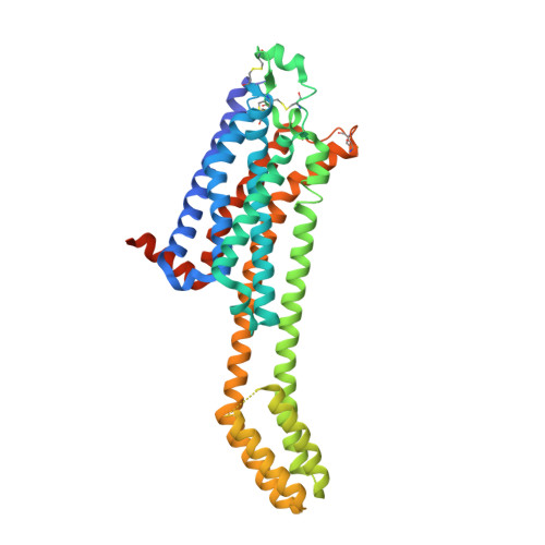
    In this study, we determined the crystal structure of an engineered human adenosine A 2A receptor bound to a partial agonist and compared it to structures cocrystallized with either a full agonist or an antagonist/inverse agonist. The interaction between the partial agonist, belonging to a class of dicyanopyridines, and amino acids in the ligand binding pocket inspired us to develop a small library of derivatives and assess their affinity in radioligand binding studies and potency and intrinsic activity in a functional, label-free, intact cell assay. It appeared that some of the derivatives retained the partial agonist profile, whereas other ligands turned into inverse agonists. We rationalized this remarkable behavior with additional computational docking studies.


  • Organizational Affiliation

    Division of Drug Discovery and Safety, Leiden Academic Centre for Drug Research, Leiden University, Einsteinweg 55, 2333 CC Leiden, The Netherlands.


Macromolecules
Find similar proteins by:  (by identity cutoff)  |  3D Structure
Entity ID: 1
MoleculeChains Sequence LengthOrganismDetailsImage
Adenosine receptor A2a,Soluble cytochrome b562,Adenosine receptor A2a433Homo sapiensEscherichia coliMutation(s): 0 
Gene Names: ADORA2AADORA2cybC
Membrane Entity: Yes 
UniProt & NIH Common Fund Data Resources
Find proteins for P0ABE7 (Escherichia coli)
Explore P0ABE7 
Go to UniProtKB:  P0ABE7
Find proteins for P29274 (Homo sapiens)
Explore P29274 
Go to UniProtKB:  P29274
PHAROS:  P29274
GTEx:  ENSG00000128271 
Entity Groups  
Sequence Clusters30% Identity50% Identity70% Identity90% Identity95% Identity100% Identity
UniProt GroupsP0ABE7P29274
Sequence Annotations
Expand
  • Reference Sequence
Small Molecules
Ligands 4 Unique
IDChains Name / Formula / InChI Key2D Diagram3D Interactions
CLR
Query on CLR

Download Ideal Coordinates CCD File 
G [auth A]CHOLESTEROL
C27 H46 O
HVYWMOMLDIMFJA-DPAQBDIFSA-N
RVZ (Subject of Investigation/LOI)
Query on RVZ

Download Ideal Coordinates CCD File 
B [auth A]2-azanyl-6-(1~{H}-imidazol-2-ylmethylsulfanyl)-4-phenyl-pyridine-3,5-dicarbonitrile
C17 H12 N6 S
MUJWBVGOOQHTTJ-UHFFFAOYSA-N
OLA
Query on OLA

Download Ideal Coordinates CCD File 
C [auth A],
D [auth A],
E [auth A],
F [auth A]
OLEIC ACID
C18 H34 O2
ZQPPMHVWECSIRJ-KTKRTIGZSA-N
NA
Query on NA

Download Ideal Coordinates CCD File 
H [auth A]SODIUM ION
Na
FKNQFGJONOIPTF-UHFFFAOYSA-N
Experimental Data & Validation

Experimental Data

  • Method: X-RAY DIFFRACTION
  • Resolution: 3.12 Å
  • R-Value Free: 0.255 
  • R-Value Work: 0.223 
  • R-Value Observed: 0.225 
  • Space Group: C 2 2 21
Unit Cell:
Length ( Å )Angle ( ˚ )
a = 39.541α = 90
b = 181.027β = 90
c = 140.853γ = 90
Software Package:
Software NamePurpose
BUSTERrefinement
XDSdata reduction
Aimlessdata scaling

Structure Validation

View Full Validation Report


Ligand Structure Quality Assessment 


Entry History 

Revision History  (Full details and data files)

  • Version 1.0: 2021-04-07
    Type: Initial release
  • Version 1.1: 2021-04-21
    Changes: Database references
  • Version 1.2: 2024-01-31
    Changes: Data collection, Database references, Refinement description