7AKD | pdb_00007akd

Structure of the SARS-CoV-2 spike glycoprotein in complex with the 47D11 neutralizing antibody Fab fragment


Experimental Data Snapshot

  • Method: ELECTRON MICROSCOPY
  • Resolution: 4.00 Å
  • Aggregation State: PARTICLE 
  • Reconstruction Method: SINGLE PARTICLE 

wwPDB Validation 3D Report Full Report

Validation slider image for 7AKD

This is version 1.2 of the entry. See complete history

Literature

Structural insights into the cross-neutralization of SARS-CoV and SARS-CoV-2 by the human monoclonal antibody 47D11.

Fedry, J.Hurdiss, D.L.Wang, C.Li, W.Obal, G.Drulyte, I.Du, W.Howes, S.C.van Kuppeveld, F.J.M.Forster, F.Bosch, B.J.

(2021) Sci Adv 7

  • DOI: https://doi.org/10.1126/sciadv.abf5632
  • Primary Citation Related Structures: 
    7AKD, 7AKJ

  • PubMed Abstract: 

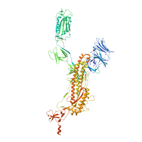
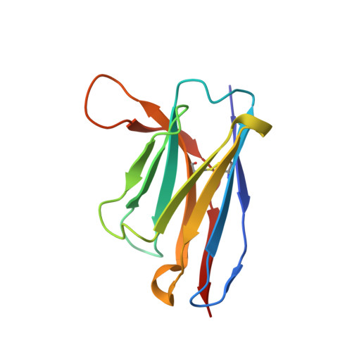
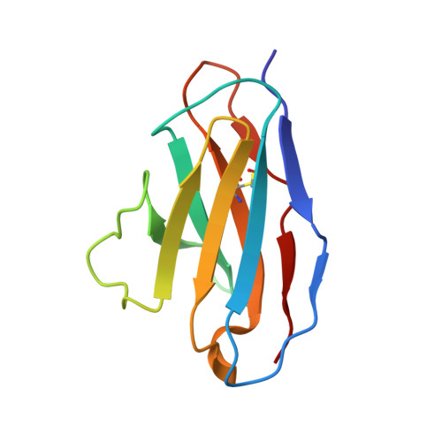
    The emergence of SARS-CoV-2 antibody escape mutations highlights the urgent need for broadly neutralizing therapeutics. We previously identified a human monoclonal antibody, 47D11, capable of cross-neutralizing SARS-CoV-2 and SARS-CoV and protecting against the associated respiratory disease in an animal model. Here, we report cryo-EM structures of both trimeric spike ectodomains in complex with the 47D11 Fab. 47D11 binds to the closed receptor-binding domain, distal to the ACE2 binding site. The CDRL3 stabilizes the N343 glycan in an upright conformation, exposing a mutationally constrained hydrophobic pocket, into which the CDRH3 loop inserts two aromatic residues. 47D11 stabilizes a partially open conformation of the SARS-CoV-2 spike, suggesting that it could be used effectively in combination with other antibodies targeting the exposed receptor-binding motif. Together, these results reveal a cross-protective epitope on the SARS-CoV-2 spike and provide a structural roadmap for the development of 47D11 as a prophylactic or postexposure therapy for COVID-19.


  • Organizational Affiliation
    • Cryo-Electron Microscopy, Bijvoet Center for Biomolecular Research, Department of Chemistry, Faculty of Science, Utrecht University, Padualaan 8, 3584 CH Utrecht, Netherlands.

Macromolecule Content 

  • Total Structure Weight: 490.25 kDa 
  • Atom Count: 27,358 
  • Modeled Residue Count: 3,404 
  • Deposited Residue Count: 4,312 
  • Unique protein chains: 3

Macromolecules

Find similar proteins by:|  3D Structure
Entity ID: 1
MoleculeChains  Sequence LengthOrganismDetailsImage
Spike glycoprotein
A, B, C
1,286Severe acute respiratory syndrome coronavirus 2Mutation(s): 0 
Gene Names: S2
UniProt
Find proteins for P0DTC2 (Severe acute respiratory syndrome coronavirus 2)
Explore P0DTC2 
Go to UniProtKB:  P0DTC2
Entity Groups
Sequence Clusters30% Identity50% Identity70% Identity90% Identity95% Identity100% Identity
UniProt GroupP0DTC2
Glycosylation
Glycosylation Sites: 6Go to GlyGen: P0DTC2-1
Sequence Annotations
Expand
Reference Sequence
Find similar proteins by:|  3D Structure
Entity ID: 2
MoleculeChains  Sequence LengthOrganismDetailsImage
47D11 neutralizing antibody heavy chainD,
F [auth H]
121Homo sapiensMutation(s): 0 
Find similar proteins by:|  3D Structure
Entity ID: 3
MoleculeChains  Sequence LengthOrganismDetailsImage
47D11 neutralizing antibody light chainE,
G [auth L]
106Homo sapiensMutation(s): 0 

Oligosaccharides

Help  
Entity ID: 4
MoleculeChains Length2D Diagram GlycosylationD Interactions
2-acetamido-2-deoxy-beta-D-glucopyranose-(1-4)-2-acetamido-2-deoxy-beta-D-glucopyranose
H [auth F],
K [auth J],
L [auth K],
M,
P,
H [auth F],
K [auth J],
L [auth K],
M,
P,
Q,
R,
V,
W,
X
2N-Glycosylation
Glycosylation Resources
GlyTouCan: G42666HT
GlyCosmos: G42666HT
GlyGen: G42666HT
Entity ID: 5
MoleculeChains Length2D Diagram GlycosylationD Interactions
beta-D-mannopyranose-(1-4)-2-acetamido-2-deoxy-beta-D-glucopyranose-(1-4)-2-acetamido-2-deoxy-beta-D-glucopyranoseI [auth G],
J [auth I],
N
3N-Glycosylation
Glycosylation Resources
GlyTouCan: G15407YE
GlyCosmos: G15407YE
GlyGen: G15407YE
Entity ID: 6
MoleculeChains Length2D Diagram GlycosylationD Interactions
alpha-D-mannopyranose-(1-3)-[alpha-D-mannopyranose-(3-6)]beta-D-mannopyranose-(1-4)-2-acetamido-2-deoxy-beta-D-glucopyranose-(1-4)-2-acetamido-2-deoxy-beta-D-glucopyranose
O
5N/AN-Glycosylation
Entity ID: 7
MoleculeChains Length2D Diagram GlycosylationD Interactions
beta-D-mannopyranose-(1-4)-2-acetamido-2-deoxy-beta-D-glucopyranose-(1-4)-[alpha-L-fucopyranose-(1-6)]2-acetamido-2-deoxy-beta-D-glucopyranose
S
4N-Glycosylation
Glycosylation Resources
GlyTouCan: G32152BH
GlyCosmos: G32152BH
GlyGen: G32152BH
Entity ID: 8
MoleculeChains Length2D Diagram GlycosylationD Interactions
alpha-D-mannopyranose-(1-3)-alpha-D-mannopyranose-(1-6)-beta-D-mannopyranose-(1-4)-2-acetamido-2-deoxy-beta-D-glucopyranose-(1-4)-[alpha-L-fucopyranose-(1-6)]2-acetamido-2-deoxy-beta-D-glucopyranose
T
6N-Glycosylation
Glycosylation Resources
GlyTouCan: G29616NS
GlyCosmos: G29616NS
GlyGen: G29616NS
Entity ID: 9
MoleculeChains Length2D Diagram GlycosylationD Interactions
alpha-D-mannopyranose-(1-6)-alpha-D-mannopyranose
U
2N/A
Glycosylation Resources
GlyTouCan: G77384RA
GlyCosmos: G77384RA
GlyGen: G77384RA

Small Molecules

Ligands 3 Unique
IDChains Name / Formula / InChI Key2D Diagram3D Interactions
NAG

Query on NAG


Download:Ideal Coordinates CCD File
AA [auth A]
AB [auth C]
BA [auth A]
BB [auth C]
CA [auth A]
AA [auth A],
AB [auth C],
BA [auth A],
BB [auth C],
CA [auth A],
DA [auth A],
EA [auth A],
FA [auth A],
GA [auth A],
IA [auth B],
JA [auth B],
KA [auth B],
LA [auth B],
MA [auth B],
OA [auth B],
PA [auth B],
QA [auth B],
RA [auth B],
SA [auth B],
TA [auth B],
UA [auth B],
VA [auth C],
WA [auth C],
XA [auth C],
Y [auth A],
YA [auth C],
Z [auth A],
ZA [auth C]
2-acetamido-2-deoxy-beta-D-glucopyranose
C8 H15 N O6
OVRNDRQMDRJTHS-FMDGEEDCSA-N
MAN

Query on MAN


Download:Ideal Coordinates CCD File
DB [auth D],
EB [auth H]
alpha-D-mannopyranose
C6 H12 O6
WQZGKKKJIJFFOK-PQMKYFCFSA-N
FUC

Query on FUC


Download:Ideal Coordinates CCD File
CB [auth C],
HA [auth A],
NA [auth B]
alpha-L-fucopyranose
C6 H12 O5
SHZGCJCMOBCMKK-SXUWKVJYSA-N

Experimental Data & Validation

Experimental Data

  • Method: ELECTRON MICROSCOPY
  • Resolution: 4.00 Å
  • Aggregation State: PARTICLE 
  • Reconstruction Method: SINGLE PARTICLE 
EM Software:
TaskSoftware PackageVersion
RECONSTRUCTIONRELION

Structure Validation

View Full Validation Report


Entry History 

& Funding Information

Deposition Data


Funding OrganizationLocationGrant Number
European Research Council (ERC)Netherlands724425-BENDER
H2020 Marie Curie Actions of the European CommissionNetherlands792575
H2020 Marie Curie Actions of the European CommissionNetherlands842333
European Molecular Biology Organization (EMBO)NetherlandsALTF-948-2017
European Molecular Biology Organization (EMBO)NetherlandsALTF-1172-2018

Revision History  (Full details and data files)

  • Version 1.0: 2021-05-19
    Type: Initial release
  • Version 1.1: 2021-06-16
    Changes: Database references
  • Version 1.2: 2024-11-06
    Changes: Data collection, Database references, Structure summary