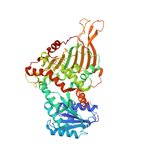
7ZVD

K89 acetylated glucose-6-phosphate dehydrogenase (G6PD) in a complex with structural NADP+


Experimental Data Snapshot

  • Method: X-RAY DIFFRACTION
  • Resolution: 2.46 Å
  • R-Value Free: 0.289 
  • R-Value Work: 0.208 
  • R-Value Observed: 0.212 

wwPDB Validation   3D Report Full Report


Ligand Structure Quality Assessment 


This is version 1.3 of the entry. See complete history


Literature

Acetylation-dependent coupling between G6PD activity and apoptotic signaling.

Wu, F.Muskat, N.H.Dvilansky, I.Koren, O.Shahar, A.Gazit, R.Elia, N.Arbely, E.

(2023) Nat Commun 14: 6208-6208

  • DOI: https://doi.org/10.1038/s41467-023-41895-2
  • Primary Citation of Related Structures:  
    7ZVD, 7ZVE

  • PubMed Abstract: 

    Lysine acetylation has been discovered in thousands of non-histone human proteins, including most metabolic enzymes. Deciphering the functions of acetylation is key to understanding how metabolic cues mediate metabolic enzyme regulation and cellular signaling. Glucose-6-phosphate dehydrogenase (G6PD), the rate-limiting enzyme in the pentose phosphate pathway, is acetylated on multiple lysine residues. Using site-specifically acetylated G6PD, we show that acetylation can activate (AcK89) and inhibit (AcK403) G6PD. Acetylation-dependent inactivation is explained by structural studies showing distortion of the dimeric structure and active site of G6PD. We provide evidence for acetylation-dependent K95/97 ubiquitylation of G6PD and Y503 phosphorylation, as well as interaction with p53 and induction of early apoptotic events. Notably, we found that the acetylation of a single lysine residue coordinates diverse acetylation-dependent processes. Our data provide an example of the complex roles of acetylation as a posttranslational modification that orchestrates the regulation of enzymatic activity, posttranslational modifications, and apoptotic signaling.


  • Organizational Affiliation

    Department of Chemistry, Ben-Gurion University of the Negev, Beer-Sheva, 8410501, Israel.


Macromolecules
Find similar proteins by:  (by identity cutoff)  |  3D Structure
Entity ID: 1
MoleculeChains Sequence LengthOrganismDetailsImage
Glucose-6-phosphate 1-dehydrogenaseA [auth N]484Homo sapiensMutation(s): 0 
Gene Names: G6PD
EC: 1.1.1.49
UniProt & NIH Common Fund Data Resources
Find proteins for P11413 (Homo sapiens)
Explore P11413 
Go to UniProtKB:  P11413
PHAROS:  P11413
GTEx:  ENSG00000160211 
Entity Groups  
Sequence Clusters30% Identity50% Identity70% Identity90% Identity95% Identity100% Identity
UniProt GroupP11413
Sequence Annotations
Expand
  • Reference Sequence
Small Molecules
Ligands 1 Unique
IDChains Name / Formula / InChI Key2D Diagram3D Interactions
NAP (Subject of Investigation/LOI)
Query on NAP

Download Ideal Coordinates CCD File 
B [auth N]NADP NICOTINAMIDE-ADENINE-DINUCLEOTIDE PHOSPHATE
C21 H28 N7 O17 P3
XJLXINKUBYWONI-NNYOXOHSSA-N
Modified Residues  1 Unique
IDChains TypeFormula2D DiagramParent
ALY
Query on ALY
A [auth N]L-PEPTIDE LINKINGC8 H16 N2 O3LYS
Experimental Data & Validation

Experimental Data

  • Method: X-RAY DIFFRACTION
  • Resolution: 2.46 Å
  • R-Value Free: 0.289 
  • R-Value Work: 0.208 
  • R-Value Observed: 0.212 
  • Space Group: F 2 2 2
Unit Cell:
Length ( Å )Angle ( ˚ )
a = 60.22α = 90
b = 172.22β = 90
c = 216.48γ = 90
Software Package:
Software NamePurpose
REFMACrefinement
XDSdata reduction
XDSdata scaling
PHASERphasing

Structure Validation

View Full Validation Report


Ligand Structure Quality Assessment 


Entry History & Funding Information

Deposition Data


Funding OrganizationLocationGrant Number
European Research Council (ERC)European Union678461
Israel Science FoundationIsrael1769/20

Revision History  (Full details and data files)

  • Version 1.0: 2023-08-23
    Type: Initial release
  • Version 1.1: 2023-11-15
    Changes: Data collection
  • Version 1.2: 2024-02-07
    Changes: Database references
  • Version 1.3: 2024-02-28
    Changes: Source and taxonomy