7ZA1

GPC3-Unc5D octamer structure and role in cell migration


Experimental Data Snapshot

  • Method: X-RAY DIFFRACTION
  • Resolution: 4.10 Å
  • R-Value Free: 0.365 
  • R-Value Work: 0.324 

wwPDB Validation   3D Report Full Report


This is version 1.1 of the entry. See complete history


Literature

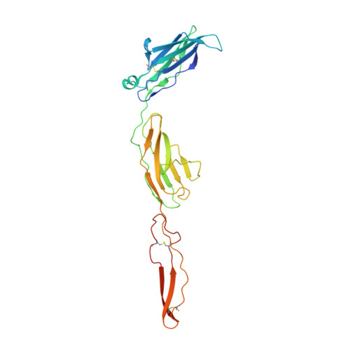
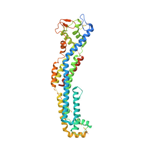
GPC3-Unc5 receptor complex structure and role in cell migration.

Akkermans, O.Delloye-Bourgeois, C.Peregrina, C.Carrasquero-Ordaz, M.Kokolaki, M.Berbeira-Santana, M.Chavent, M.Reynaud, F.Raj, R.Agirre, J.Aksu, M.White, E.S.Lowe, E.Ben Amar, D.Zaballa, S.Huo, J.Pakos, I.McCubbin, P.T.N.Comoletti, D.Owens, R.J.Robinson, C.V.Castellani, V.Del Toro, D.Seiradake, E.

(2022) Cell 185: 3931-3949.e26

  • DOI: https://doi.org/10.1016/j.cell.2022.09.025
  • Primary Citation of Related Structures:  
    7ZA1, 7ZA2, 7ZA3, 7ZAV, 7ZAW

  • PubMed Abstract: 

    Neural migration is a critical step during brain development that requires the interactions of cell-surface guidance receptors. Cancer cells often hijack these mechanisms to disseminate. Here, we reveal crystal structures of Uncoordinated-5 receptor D (Unc5D) in complex with morphogen receptor glypican-3 (GPC3), forming an octameric glycoprotein complex. In the complex, four Unc5D molecules pack into an antiparallel bundle, flanked by four GPC3 molecules. Central glycan-glycan interactions are formed by N-linked glycans emanating from GPC3 (N241 in human) and C-mannosylated tryptophans of the Unc5D thrombospondin-like domains. MD simulations, mass spectrometry and structure-based mutants validate the crystallographic data. Anti-GPC3 nanobodies enhance or weaken Unc5-GPC3 binding and, together with mutant proteins, show that Unc5/GPC3 guide migrating pyramidal neurons in the mouse cortex, and cancer cells in an embryonic xenograft neuroblastoma model. The results demonstrate a conserved structural mechanism of cell guidance, where finely balanced Unc5-GPC3 interactions regulate cell migration.


  • Organizational Affiliation

    Department of Biochemistry, University of Oxford, Oxford, UK.


Macromolecules
Find similar proteins by:  (by identity cutoff)  |  3D Structure
Entity ID: 1
MoleculeChains Sequence LengthOrganismDetailsImage
Netrin receptor UNC5DA [auth E],
B [auth F],
C [auth G],
D [auth H]
268Rattus norvegicusMutation(s): 0 
Gene Names: Unc5d
UniProt
Find proteins for F1LW30 (Rattus norvegicus)
Explore F1LW30 
Go to UniProtKB:  F1LW30
Entity Groups  
Sequence Clusters30% Identity50% Identity70% Identity90% Identity95% Identity100% Identity
UniProt GroupF1LW30
Sequence Annotations
Expand
  • Reference Sequence
Find similar proteins by:  (by identity cutoff)  |  3D Structure
Entity ID: 2
MoleculeChains Sequence LengthOrganismDetailsImage
Glypican-3E [auth A],
F [auth C],
G [auth D],
H [auth B]
464Homo sapiensMutation(s): 1 
Gene Names: GPC3OCI5
UniProt & NIH Common Fund Data Resources
Find proteins for P51654 (Homo sapiens)
Explore P51654 
Go to UniProtKB:  P51654
PHAROS:  P51654
GTEx:  ENSG00000147257 
Entity Groups  
Sequence Clusters30% Identity50% Identity70% Identity90% Identity95% Identity100% Identity
UniProt GroupP51654
Sequence Annotations
Expand
  • Reference Sequence
Oligosaccharides

Help

Entity ID: 3
MoleculeChains Length2D Diagram Glycosylation3D Interactions
2-acetamido-2-deoxy-beta-D-glucopyranose-(1-4)-2-acetamido-2-deoxy-beta-D-glucopyranose
I
2N-Glycosylation
Glycosylation Resources
GlyTouCan:  G42666HT
GlyCosmos:  G42666HT
GlyGen:  G42666HT
Entity ID: 4
MoleculeChains Length2D Diagram Glycosylation3D Interactions
alpha-D-mannopyranose-(1-3)-beta-D-mannopyranose-(1-4)-2-acetamido-2-deoxy-beta-D-glucopyranose-(1-4)-2-acetamido-2-deoxy-beta-D-glucopyranoseJ,
K,
L [auth M],
M [auth N]
4N-Glycosylation
Glycosylation Resources
GlyTouCan:  G81315DD
GlyCosmos:  G81315DD
GlyGen:  G81315DD
Small Molecules
Ligands 2 Unique
IDChains Name / Formula / InChI Key2D Diagram3D Interactions
NAG
Query on NAG

Download Ideal Coordinates CCD File 
AA [auth A]
BA [auth C]
CA [auth D]
DA [auth B]
EA [auth B]
AA [auth A],
BA [auth C],
CA [auth D],
DA [auth B],
EA [auth B],
N [auth E],
O [auth E],
R [auth F],
U [auth F],
V [auth H],
Y [auth H],
Z [auth A]
2-acetamido-2-deoxy-beta-D-glucopyranose
C8 H15 N O6
OVRNDRQMDRJTHS-FMDGEEDCSA-N
MAN
Query on MAN

Download Ideal Coordinates CCD File 
P [auth E]
Q [auth E]
S [auth F]
T [auth F]
W [auth H]
P [auth E],
Q [auth E],
S [auth F],
T [auth F],
W [auth H],
X [auth H]
alpha-D-mannopyranose
C6 H12 O6
WQZGKKKJIJFFOK-PQMKYFCFSA-N
Experimental Data & Validation

Experimental Data

  • Method: X-RAY DIFFRACTION
  • Resolution: 4.10 Å
  • R-Value Free: 0.365 
  • R-Value Work: 0.324 
  • Space Group: P 1 21 1
Unit Cell:
Length ( Å )Angle ( ˚ )
a = 102.643α = 90
b = 157.584β = 102.949
c = 126.601γ = 90
Software Package:
Software NamePurpose
REFMACrefinement
autoPROCdata reduction
xia2data scaling
PHENIXphasing

Structure Validation

View Full Validation Report


Entry History & Funding Information

Deposition Data


Funding OrganizationLocationGrant Number
Wellcome TrustUnited Kingdom202827/Z/16/Z

Revision History  (Full details and data files)

  • Version 1.0: 2022-11-02
    Type: Initial release
  • Version 1.1: 2024-01-31
    Changes: Data collection, Refinement description