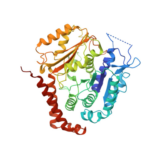
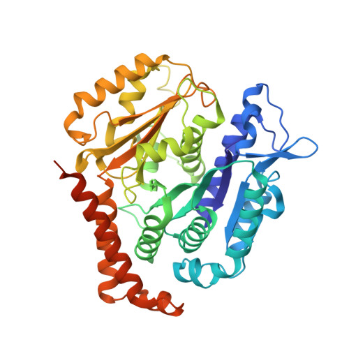
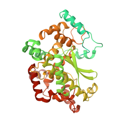
7Z6S

MATCAP bound to a human 14 protofilament microtubule


Experimental Data Snapshot

  • Method: ELECTRON MICROSCOPY
  • Resolution: 2.90 Å
  • Aggregation State: HELICAL ARRAY 
  • Reconstruction Method: SINGLE PARTICLE 

Starting Models: experimental
View more details

wwPDB Validation   3D Report Full Report


This is version 1.2 of the entry. See complete history


Literature

Posttranslational modification of microtubules by the MATCAP detyrosinase.

Landskron, L.Bak, J.Adamopoulos, A.Kaplani, K.Moraiti, M.van den Hengel, L.G.Song, J.Y.Bleijerveld, O.B.Nieuwenhuis, J.Heidebrecht, T.Henneman, L.Moutin, M.J.Barisic, M.Taraviras, S.Perrakis, A.Brummelkamp, T.R.

(2022) Science 376: eabn6020-eabn6020

  • DOI: https://doi.org/10.1126/science.abn6020
  • Primary Citation of Related Structures:  
    7Z5G, 7Z5H, 7Z6S

  • PubMed Abstract: 

    The detyrosination-tyrosination cycle involves the removal and religation of the C-terminal tyrosine of α-tubulin and is implicated in cognitive, cardiac, and mitotic defects. The vasohibin-small vasohibin-binding protein (SVBP) complex underlies much, but not all, detyrosination. We used haploid genetic screens to identify an unannotated protein, microtubule associated tyrosine carboxypeptidase (MATCAP), as a remaining detyrosinating enzyme. X-ray crystallography and cryo-electron microscopy structures established MATCAP's cleaving mechanism, substrate specificity, and microtubule recognition. Paradoxically, whereas abrogation of tyrosine religation is lethal in mice, codeletion of MATCAP and SVBP is not. Although viable, defective detyrosination caused microcephaly, associated with proliferative defects during neurogenesis, and abnormal behavior. Thus, MATCAP is a missing component of the detyrosination-tyrosination cycle, revealing the importance of this modification in brain formation.


  • Organizational Affiliation

    Oncode Institute, Division of Biochemistry, Netherlands Cancer Institute, 1066CX Amsterdam, Netherlands.


Macromolecules
Find similar proteins by:  (by identity cutoff)  |  3D Structure
Entity ID: 1
MoleculeChains Sequence LengthOrganismDetailsImage
Tubulin alpha-1B chainA,
D [auth K]
457Homo sapiensMutation(s): 0 
EC: 3.6.5
UniProt & NIH Common Fund Data Resources
Find proteins for P68363 (Homo sapiens)
Explore P68363 
Go to UniProtKB:  P68363
PHAROS:  P68363
GTEx:  ENSG00000123416 
Entity Groups  
Sequence Clusters30% Identity50% Identity70% Identity90% Identity95% Identity100% Identity
UniProt GroupP68363
Sequence Annotations
Expand
  • Reference Sequence
Find similar proteins by:  (by identity cutoff)  |  3D Structure
Entity ID: 2
MoleculeChains Sequence LengthOrganismDetailsImage
Tubulin beta-3 chainB,
C [auth H]
456Homo sapiensMutation(s): 0 
Gene Names: TUBB3TUBB4
UniProt & NIH Common Fund Data Resources
Find proteins for Q13509 (Homo sapiens)
Explore Q13509 
Go to UniProtKB:  Q13509
PHAROS:  Q13509
GTEx:  ENSG00000258947 
Entity Groups  
Sequence Clusters30% Identity50% Identity70% Identity90% Identity95% Identity100% Identity
UniProt GroupQ13509
Sequence Annotations
Expand
  • Reference Sequence
Find similar proteins by:  (by identity cutoff)  |  3D Structure
Entity ID: 3
MoleculeChains Sequence LengthOrganismDetailsImage
Uncharacterized protein KIAA0895-likeE [auth C],
F [auth E]
471Homo sapiensMutation(s): 1 
Gene Names: KIAA0895L
EC: 3.4.17.17
UniProt & NIH Common Fund Data Resources
Find proteins for Q68EN5 (Homo sapiens)
Explore Q68EN5 
Go to UniProtKB:  Q68EN5
PHAROS:  Q68EN5
GTEx:  ENSG00000196123 
Entity Groups  
Sequence Clusters30% Identity50% Identity70% Identity90% Identity95% Identity100% Identity
UniProt GroupQ68EN5
Sequence Annotations
Expand
  • Reference Sequence
Small Molecules
Ligands 4 Unique
IDChains Name / Formula / InChI Key2D Diagram3D Interactions
GTP (Subject of Investigation/LOI)
Query on GTP

Download Ideal Coordinates CCD File 
G [auth A],
K
GUANOSINE-5'-TRIPHOSPHATE
C10 H16 N5 O14 P3
XKMLYUALXHKNFT-UUOKFMHZSA-N
GDP (Subject of Investigation/LOI)
Query on GDP

Download Ideal Coordinates CCD File 
I [auth B],
J [auth H]
GUANOSINE-5'-DIPHOSPHATE
C10 H15 N5 O11 P2
QGWNDRXFNXRZMB-UUOKFMHZSA-N
ZN (Subject of Investigation/LOI)
Query on ZN

Download Ideal Coordinates CCD File 
M [auth C],
N [auth E]
ZINC ION
Zn
PTFCDOFLOPIGGS-UHFFFAOYSA-N
MG (Subject of Investigation/LOI)
Query on MG

Download Ideal Coordinates CCD File 
H [auth A],
L [auth K]
MAGNESIUM ION
Mg
JLVVSXFLKOJNIY-UHFFFAOYSA-N
Experimental Data & Validation

Experimental Data

  • Method: ELECTRON MICROSCOPY
  • Resolution: 2.90 Å
  • Aggregation State: HELICAL ARRAY 
  • Reconstruction Method: SINGLE PARTICLE 

Structure Validation

View Full Validation Report


Entry History & Funding Information

Deposition Data


Funding OrganizationLocationGrant Number
Oncode InstituteNetherlands--

Revision History  (Full details and data files)

  • Version 1.0: 2022-05-11
    Type: Initial release
  • Version 1.1: 2022-06-01
    Changes: Database references
  • Version 1.2: 2024-10-16
    Changes: Data collection, Refinement description, Structure summary