


Crystal Structures of two novel recombinases RecA
Freitag-Pohl, S., Pohl, E.To be published.
Experimental Data Snapshot
Starting Model: experimental
View more details
wwPDB Validation 3D Report Full Report

Entity ID: 1 | |||||
|---|---|---|---|---|---|
| Molecule | Chains | Sequence Length | Organism | Details | Image |
| recombinase | A [auth AAA] | 373 | unidentified | Mutation(s): 0 | 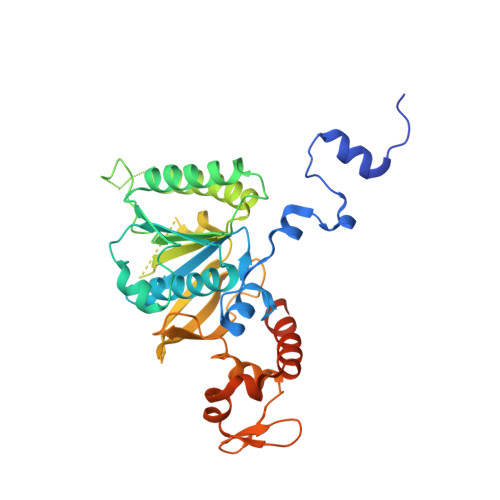 |
| Ligands 1 Unique | |||||
|---|---|---|---|---|---|
| ID | Chains | Name / Formula / InChI Key | 2D Diagram | 3D Interactions | |
| SO4 Download:Ideal Coordinates CCD File | B [auth AAA] | SULFATE ION O4 S QAOWNCQODCNURD-UHFFFAOYSA-L |  | ||
| Length ( Å ) | Angle ( ˚ ) |
|---|---|
| a = 90.895 | α = 90 |
| b = 90.895 | β = 90 |
| c = 101.438 | γ = 120 |
| Software Name | Purpose |
|---|---|
| REFMAC | refinement |
| XDS | data reduction |
| XSCALE | data scaling |
| PHASER | phasing |
| Arcimboldo | phasing |
| BUCCANEER | model building |
| Funding Organization | Location | Grant Number |
|---|---|---|
| European Union (EU) | European Union | 685778 |


