


Crystal structure of PEX13 SH3 domain of Trypanosoma cruzi
Sonani, R.R., Dubin, G.To be published.
Experimental Data Snapshot
Starting Model: experimental
View more details
wwPDB Validation 3D Report Full Report

Entity ID: 1 | |||||
|---|---|---|---|---|---|
| Molecule | Chains | Sequence Length | Organism | Details | Image |
| Putative peroxin 13 | 81 | Trypanosoma cruzi | Mutation(s): 0 Gene Names: C4B63_4g75 | 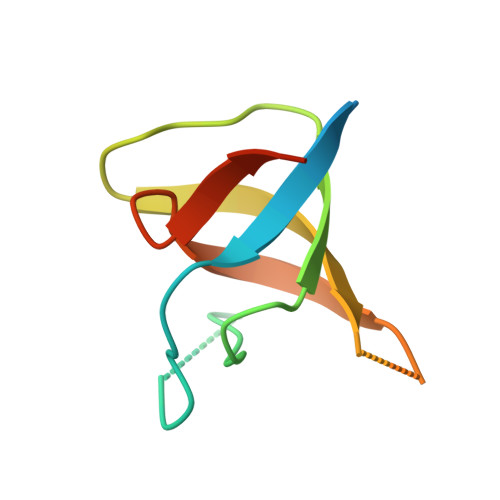 | |
UniProt | |||||
Entity Groups | |||||
| Sequence Clusters | 30% Identity50% Identity70% Identity90% Identity95% Identity100% Identity | ||||
| UniProt Group | A0A2V2VYH2 | ||||
Sequence AnnotationsExpand | |||||
Reference Sequence | |||||
| Length ( Å ) | Angle ( ˚ ) |
|---|---|
| a = 52.481 | α = 90 |
| b = 52.481 | β = 90 |
| c = 113.969 | γ = 120 |
| Software Name | Purpose |
|---|---|
| REFMAC | refinement |
| PDB_EXTRACT | data extraction |
| XDS | data reduction |
| Aimless | data scaling |
| PHASER | phasing |
| Funding Organization | Location | Grant Number |
|---|---|---|
| Polish National Science Centre | Poland | 2017/26/M/NZ1/00797 |
| Foundation for Polish Science | Poland | TEAM TECH CORE FACILITY/2017-4/6 |


