7Z2R | pdb_00007z2r

Differences between the GluD1 and GluD2 receptors revealed by GluD1 X-ray crystallography, binding studies and molecular dynamics


Experimental Data Snapshot

  • Method: X-RAY DIFFRACTION
  • Resolution: 2.57 Å
  • R-Value Free: 
    0.252 (Depositor), 0.250 (DCC) 
  • R-Value Work: 
    0.215 (Depositor), 0.218 (DCC) 
  • R-Value Observed: 
    0.217 (Depositor) 

Starting Model: experimental
View more details

wwPDB Validation 3D Report Full Report

Validation slider image for 7Z2R

This is version 1.2 of the entry. See complete history

Literature

Differences between the GluD1 and GluD2 receptors revealed by GluD1 X-ray crystallography, binding studies and molecular dynamics.

Masternak, M.Koch, A.Laulumaa, S.Tapken, D.Hollmann, M.Jorgensen, F.S.Kastrup, J.S.

(2023) FEBS J 290: 3781-3801

  • DOI: https://doi.org/10.1111/febs.16631
  • Primary Citation Related Structures: 
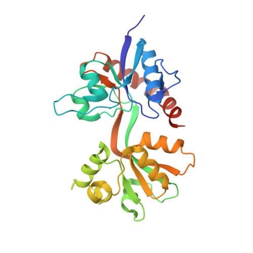
    7Z2R

  • PubMed Abstract: 

    Ionotropic glutamate receptors are ligand-gated ion channels essential for fast excitatory neurotransmission in the brain. In contrast to most other members of the iGluR family, the subfamily of delta receptors, GluD1 and GluD2, does not bind glutamate but glycine/D-serine. GluD1 is widely expressed in the brain and the inner ear, where it is required for high-frequency hearing. Furthermore, it has been associated with schizophrenia, autism and depression. X-ray structures of the ligand-binding domain (LBD) of GluD2 have been published; however, no high-resolution structure is available for the ligand-binding domain of GluD1 (GluD1-LBD). Here, we report the X-ray crystal structure of the GluD1-LBD in its apo form at 2.57 Å resolution. Using isothermal titration calorimetry, we show that D-serine binds to the GluD1-LBD in an exothermic manner with a K d of 160 μm, which is approximately five-fold greater than at GluD2. Furthermore, we identify Glu822 as a critical determinant of receptor activation in GluD1 A654T. In contrast to studies on the GluD2 lurcher mutant A654T, we did not observe any effect of 1 mm D-serine on the spontaneous currents at mouse GluD1 A654T by electrophysiological recordings of Xenopus laevis oocytes as previously also reported by others. These results point towards differences in the structure and dynamics between GluD1 and GluD2. Molecular dynamics simulations were employed to address this observation, suggesting that the apo structure of GluD1 is less flexible than the apo structure of GluD2 and that Pro725 in GluD1 may affect the interlobe closure of the ligand-binding domain of GluD1.


  • Organizational Affiliation
    • Department of Drug Design and Pharmacology, University of Copenhagen, Denmark.

Macromolecule Content 

  • Total Structure Weight: 59.86 kDa 
  • Atom Count: 4,187 
  • Modeled Residue Count: 517 
  • Deposited Residue Count: 532 
  • Unique protein chains: 1

Macromolecules

Find similar proteins by:|  3D Structure
Entity ID: 1
MoleculeChains  Sequence LengthOrganismDetailsImage
Glutamate receptor ionotropic, delta-1
A, B
266Rattus norvegicusMutation(s): 0 
Gene Names: Grid1
UniProt
Find proteins for Q62640 (Rattus norvegicus)
Explore Q62640 
Go to UniProtKB:  Q62640
Entity Groups
Sequence Clusters30% Identity50% Identity70% Identity90% Identity95% Identity100% Identity
UniProt GroupQ62640
Sequence Annotations
Expand
Reference Sequence

Experimental Data & Validation

Experimental Data

  • Method: X-RAY DIFFRACTION
  • Resolution: 2.57 Å
  • R-Value Free:  0.252 (Depositor), 0.250 (DCC) 
  • R-Value Work:  0.215 (Depositor), 0.218 (DCC) 
  • R-Value Observed: 0.217 (Depositor) 
Space Group: P 1 21 1
Unit Cell:
Length ( Å )Angle ( ˚ )
a = 33.31α = 90
b = 136.44β = 96.98
c = 54.61γ = 90
Software Package:
Software NamePurpose
PHENIXrefinement
PDB_EXTRACTdata extraction
AutoProcessdata processing
XSCALEdata scaling
PHASERphasing
TRUNCATEdata reduction

Structure Validation

View Full Validation Report


Entry History 

& Funding Information

Deposition Data


Funding OrganizationLocationGrant Number
Danish Council for Independent ResearchDenmark--

Revision History  (Full details and data files)

  • Version 1.0: 2023-01-11
    Type: Initial release
  • Version 1.1: 2023-08-16
    Changes: Database references, Refinement description
  • Version 1.2: 2024-01-31
    Changes: Data collection, Refinement description