Crystal structure and functional characterization of a novel bacterial lignin-degrading dye-decolorizing peroxidase.
Catucci, G., Zhang, C., Pernaci, A., Cappa, F., Sadeghi, S.J., Di Nardo, G., Gilardi, G.(2025) Int J Biol Macromol 297: 139900-139900
- PubMed: 39818373 Search on PubMed
- DOI: https://doi.org/10.1016/j.ijbiomac.2025.139900
- Primary Citation Related Structures:
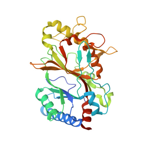
7YZT - PubMed Abstract:
A new gene coding for an iron-containing enzyme was identified in the genome of Acinetobacter radioresistens. Bioinformatics analysis allowed the assignment of the protein to DyP peroxidases, due to the presence of conserved residues involved in heme binding and catalysis. Moreover, Ar-DyP is located in an operon coding also for other enzymes involved in iron uptake and regulation. The crystal structure of Ar-DyP determined at 1.85 Å resolution shows that the heme pocket Ar-DyP is "wet" forming a continuous hydrogen-bond network that enables the communication between heme and distal residues. Moreover, as shown by the crystal structure and covalent crosslinking experiments, Ar-DyP uses a long-range electron transfer pathway involving His-181 and Tyr-241, in the active site and on the surface of the enzyme, respectively. This pathway allows oxidation of substrates of different sizes, including Kraft lignin. Indeed, the biochemical characterization showed that Ar-Dyp oxidizes ABTS and Reactive Blue 19 (turnover numbers of 500 and 464 min -1 , respectively), but also phenolic compounds such as guaiacol and pyrogallol (turnover numbers of 7.4 and 1.8 min -1 respectively). Overall, the data shows that Ar-DyP is a promising candidate for applications in lignin valorization, bioremediation and industrial processes involving the breakdown of phenolic compounds.
- Department of Life Sciences and Systems Biology, University of Torino, Italy.
Organizational Affiliation: