7YZ8

Triclinic crystal structure of YTHDF1 YTH domain (544AVV546 mutant)


Experimental Data Snapshot

  • Method: X-RAY DIFFRACTION
  • Resolution: 2.50 Å
  • R-Value Free: 0.243 
  • R-Value Work: 0.184 
  • R-Value Observed: 0.187 

wwPDB Validation   3D Report Full Report


This is version 1.2 of the entry. See complete history


Literature

Pliability in the m 6 A-Binding Region Extends Druggability of YTH Domains.

Cazzanelli, G.Dalle Vedove, A.Spagnolli, G.Terruzzi, L.Colasurdo, E.Boldrini, A.Patsilinakos, A.Sturlese, M.Grottesi, A.Biasini, E.Provenzani, A.Quattrone, A.Lolli, G.

(2024) J Chem Inf Model 

  • DOI: https://doi.org/10.1021/acs.jcim.4c00051
  • Primary Citation of Related Structures:  
    7YYE, 7YYF, 7YYJ, 7YZ8

  • PubMed Abstract: 

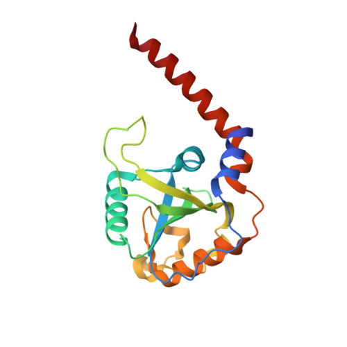
    Epitranscriptomic mRNA modifications affect gene expression, with their altered balance detected in various cancers. YTHDF proteins contain the YTH reader domain recognizing the m 6 A mark on mRNA and represent valuable drug targets. Crystallographic structures have been determined for all three family members; however, discrepancies are present in the organization of the m 6 A-binding pocket. Here, we present new crystallographic structures of the YTH domain of YTHDF1, accompanied by computational studies, showing that this domain can exist in different stable conformations separated by a significant energetic barrier. During the transition, additional conformations are explored, with peculiar druggable pockets appearing and offering new opportunities for the design of YTH-interfering small molecules.


  • Organizational Affiliation

    Department of Cellular, Computational and Integrative Biology─CIBIO, University of Trento, via Sommarive 9, 38123 Povo, Trento, Italy.


Macromolecules
Find similar proteins by:  (by identity cutoff)  |  3D Structure
Entity ID: 1
MoleculeChains Sequence LengthOrganismDetailsImage
YTH domain-containing family protein 1
A, B, C, D, E
A, B, C, D, E, F, G, H, I, J, K, L
200Homo sapiensMutation(s): 3 
Gene Names: YTHDF1C20orf21
UniProt & NIH Common Fund Data Resources
Find proteins for Q9BYJ9 (Homo sapiens)
Explore Q9BYJ9 
Go to UniProtKB:  Q9BYJ9
PHAROS:  Q9BYJ9
GTEx:  ENSG00000149658 
Entity Groups  
Sequence Clusters30% Identity50% Identity70% Identity90% Identity95% Identity100% Identity
UniProt GroupQ9BYJ9
Sequence Annotations
Expand
  • Reference Sequence
Small Molecules
Ligands 1 Unique
IDChains Name / Formula / InChI Key2D Diagram3D Interactions
SCN
Query on SCN

Download Ideal Coordinates CCD File 
M [auth A]
N [auth B]
O [auth C]
P [auth D]
Q [auth E]
M [auth A],
N [auth B],
O [auth C],
P [auth D],
Q [auth E],
R [auth F],
S [auth G],
T [auth H],
U [auth I],
V [auth J],
W [auth K],
X [auth L]
THIOCYANATE ION
C N S
ZMZDMBWJUHKJPS-UHFFFAOYSA-M
Experimental Data & Validation

Experimental Data

  • Method: X-RAY DIFFRACTION
  • Resolution: 2.50 Å
  • R-Value Free: 0.243 
  • R-Value Work: 0.184 
  • R-Value Observed: 0.187 
  • Space Group: P 1
Unit Cell:
Length ( Å )Angle ( ˚ )
a = 71.225α = 72.87
b = 92.571β = 79.05
c = 113.94γ = 67.87
Software Package:
Software NamePurpose
PHENIXrefinement
XDSdata reduction
Aimlessdata scaling
PHASERphasing
PDB_EXTRACTdata extraction

Structure Validation

View Full Validation Report


Entry History & Funding Information

Deposition Data


Funding OrganizationLocationGrant Number
Italian Association for Cancer ResearchItalyMFAG 2017 - ID. 19882

Revision History  (Full details and data files)

  • Version 1.0: 2023-03-01
    Type: Initial release
  • Version 1.1: 2024-02-07
    Changes: Data collection, Refinement description
  • Version 1.2: 2024-03-13
    Changes: Database references