7YWY | pdb_00007ywy

Structure of the GroEL chaperonin in complex with the CnoX chaperedoxin


Experimental Data Snapshot

  • Method: ELECTRON MICROSCOPY
  • Resolution: 3.40 Å
  • Aggregation State: PARTICLE 
  • Reconstruction Method: SINGLE PARTICLE 

Starting Models: experimental
View more details

wwPDB Validation   3D Report Full Report


This is version 1.2 of the entry. See complete history


Literature

A molecular device for the redox quality control of GroEL/ES substrates.

Dupuy, E.Van der Verren, S.E.Lin, J.Wilson, M.A.Dachsbeck, A.V.Viela, F.Latour, E.Gennaris, A.Vertommen, D.Dufrene, Y.F.Iorga, B.I.Goemans, C.V.Remaut, H.Collet, J.F.

(2023) Cell 186: 1039-1049.e17

  • DOI: https://doi.org/10.1016/j.cell.2023.01.013
  • Primary Citation of Related Structures:  
    7YWY

  • PubMed Abstract: 

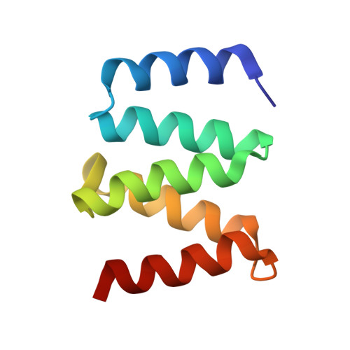
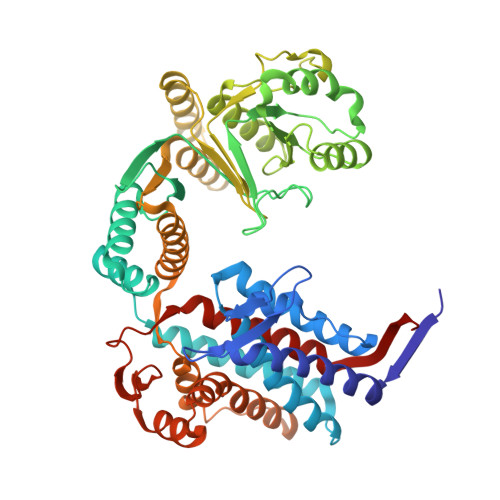
    Hsp60 chaperonins and their Hsp10 cofactors assist protein folding in all living cells, constituting the paradigmatic example of molecular chaperones. Despite extensive investigations of their structure and mechanism, crucial questions regarding how these chaperonins promote folding remain unsolved. Here, we report that the bacterial Hsp60 chaperonin GroEL forms a stable, functionally relevant complex with the chaperedoxin CnoX, a protein combining a chaperone and a redox function. Binding of GroES (Hsp10 cofactor) to GroEL induces CnoX release. Cryoelectron microscopy provided crucial structural information on the GroEL-CnoX complex, showing that CnoX binds GroEL outside the substrate-binding site via a highly conserved C-terminal α-helix. Furthermore, we identified complexes in which CnoX, bound to GroEL, forms mixed disulfides with GroEL substrates, indicating that CnoX likely functions as a redox quality-control plugin for GroEL. Proteins sharing structural features with CnoX exist in eukaryotes, suggesting that Hsp60 molecular plugins have been conserved through evolution.


  • Organizational Affiliation
    • WELBIO, Avenue Hippocrate 75, 1200 Brussels, Belgium; de Duve Institute, Université catholique de Louvain, Avenue Hippocrate 75, 1200 Brussels, Belgium.

Macromolecules
Find similar proteins by:  (by identity cutoff)  |  3D Structure
Entity ID: 1
MoleculeChains Sequence LengthOrganismDetailsImage
Chaperedoxin88Escherichia coliMutation(s): 0 
Gene Names: cnoXGHT02_06645
UniProt
Find proteins for P77395 (Escherichia coli (strain K12))
Explore P77395 
Go to UniProtKB:  P77395
Entity Groups  
Sequence Clusters30% Identity50% Identity70% Identity90% Identity95% Identity100% Identity
UniProt GroupP77395
Sequence Annotations
Expand
  • Reference Sequence
Find similar proteins by:  (by identity cutoff)  |  3D Structure
Entity ID: 2
MoleculeChains Sequence LengthOrganismDetailsImage
Chaperonin GroEL524Escherichia coliMutation(s): 0 
Gene Names: groLAMK64_004370
EC: 5.6.1.7
UniProt
Find proteins for P0A6F5 (Escherichia coli (strain K12))
Explore P0A6F5 
Go to UniProtKB:  P0A6F5
Entity Groups  
Sequence Clusters30% Identity50% Identity70% Identity90% Identity95% Identity100% Identity
UniProt GroupP0A6F5
Sequence Annotations
Expand
  • Reference Sequence
Experimental Data & Validation

Experimental Data

  • Method: ELECTRON MICROSCOPY
  • Resolution: 3.40 Å
  • Aggregation State: PARTICLE 
  • Reconstruction Method: SINGLE PARTICLE 
EM Software:
TaskSoftware PackageVersion
RECONSTRUCTIONRELION3.0
MODEL REFINEMENTPHENIX

Structure Validation

View Full Validation Report


Entry History & Funding Information

Deposition Data


Funding OrganizationLocationGrant Number
Research Foundation - Flanders (FWO)BelgiumG0G0818N
Fonds de la Recherche Scientifique (FNRS)BelgiumWELBIO-CR-2019C-03

Revision History  (Full details and data files)

  • Version 1.0: 2023-02-22
    Type: Initial release
  • Version 1.1: 2024-07-17
    Changes: Data collection, Refinement description
  • Version 1.2: 2024-09-04
    Changes: Data collection, Database references