7YQA

Crystal structure of D-threonine aldolase from Chlamydomonas reinhardtii


Experimental Data Snapshot

  • Method: X-RAY DIFFRACTION
  • Resolution: 1.85 Å
  • R-Value Free: 0.249 
  • R-Value Work: 0.217 
  • R-Value Observed: 0.219 

wwPDB Validation   3D Report Full Report


This is version 1.1 of the entry. See complete history


Literature

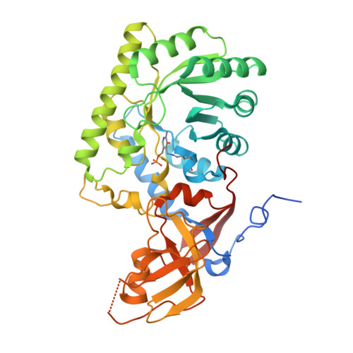
Structure of pyridoxal 5'-phosphate-bound D-threonine aldolase from Chlamydomonas reinhardtii.

Hirato, Y.Goto, M.Mizobuchi, T.Muramatsu, H.Tanigawa, M.Nishimura, K.

(2023) Acta Crystallogr F Struct Biol Commun 79: 31-37

  • DOI: https://doi.org/10.1107/S2053230X23000304
  • Primary Citation of Related Structures:  
    7YQA

  • PubMed Abstract: 

    D-Threonine aldolase (DTA) is a pyridoxal-5'-phosphate-dependent enzyme which catalyzes the reversible aldol reaction of glycine with a corresponding aldehyde to yield the D-form β-hydroxy-α-amino acid. This study produced and investigated the crystal structure of DTA from Chlamydomonas reinhardtii (CrDTA) at 1.85 Å resolution. To our knowledge, this is the first report on the crystal structure of eukaryotic DTA. Compared with the structure of bacterial DTA, CrDTA has a similar arrangement of active-site residues. On the other hand, we speculated that some non-conserved residues alter the affinity for substrates and inhibitors. The structure of CrDTA could provide insights into the structural framework for structure-guided protein engineering studies to modify reaction selectivity.


  • Organizational Affiliation

    Department of Biomolecular Science, Faculty of Science, Toho University, 2-2-1 Miyama, Funabashi, Chiba 274-8510, Japan.


Macromolecules
Find similar proteins by:  (by identity cutoff)  |  3D Structure
Entity ID: 1
MoleculeChains Sequence LengthOrganismDetailsImage
D-threonine aldolase
A, B, C, D
428Chlamydomonas reinhardtiiMutation(s): 0 
EC: 4.1.2.42
UniProt
Find proteins for A0A1C9ZZ39 (Chlamydomonas reinhardtii)
Explore A0A1C9ZZ39 
Go to UniProtKB:  A0A1C9ZZ39
Entity Groups  
Sequence Clusters30% Identity50% Identity70% Identity90% Identity95% Identity100% Identity
UniProt GroupA0A1C9ZZ39
Sequence Annotations
Expand
  • Reference Sequence
Experimental Data & Validation

Experimental Data

  • Method: X-RAY DIFFRACTION
  • Resolution: 1.85 Å
  • R-Value Free: 0.249 
  • R-Value Work: 0.217 
  • R-Value Observed: 0.219 
  • Space Group: P 1
Unit Cell:
Length ( Å )Angle ( ˚ )
a = 64.792α = 77.07
b = 74.096β = 69.34
c = 89.939γ = 71.93
Software Package:
Software NamePurpose
REFMACrefinement
XDSdata reduction
SCALAdata scaling
PDB_EXTRACTdata extraction
MOLREPphasing

Structure Validation

View Full Validation Report


Entry History & Funding Information

Deposition Data


Funding OrganizationLocationGrant Number
Not funded--

Revision History  (Full details and data files)

  • Version 1.0: 2023-02-15
    Type: Initial release
  • Version 1.1: 2023-11-29
    Changes: Data collection, Refinement description