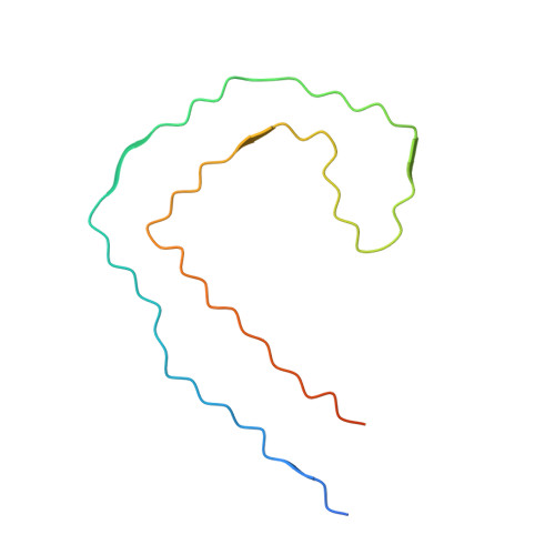
Subtle change of fibrillation condition leads to substantial alteration of recombinant Tau fibril structure.
Li, X., Zhang, S., Liu, Z., Tao, Y., Xia, W., Sun, Y., Liu, C., Le, W., Sun, B., Li, D.(2022) iScience 25: 105645-105645
- PubMed: 36505939
- DOI: https://doi.org/10.1016/j.isci.2022.105645
- Primary Citation of Related Structures:
7YMN, 7YPG - PubMed Abstract:
In vitro assembly of amyloid fibrils that recapitulate those in human brains is very useful for fundamental and applied research on the amyloid formation, pathology, and clinical detection. Recent success in the assembly of Tau fibrils in vitro enables the recapitulation of the paired helical filament (PHF) of Tau extracted from brains of patients with Alzheimer's disease (AD). However, following the protocol, we observed that Tau constructs including 297-391 and a mixture of 266-391 (3R)/297-391, which are expected to predominantly form PHF-like fibrils, form highly heterogeneous fibrils instead. Moreover, the seemingly PHF-like fibril formed by Tau 297-391 exhibits a distinctive atomic structure with a spindle-like fold, that is neither PHF-like or similar to any known Tau fibril structures revealed by cryo-electron microscopy (cryo-EM). Our work highlights the high sensitivity of amyloid fibril formation to subtle conditional changes and suggests high-resolution structural characterization to in vitro assembled fibrils prior to further laboratory use.
- Bio-X Institutes, Key Laboratory for the Genetics of Developmental and Neuropsychiatric Disorders (Ministry of Education), Shanghai Jiao Tong University, Shanghai 200030, China.
Organizational Affiliation:


