7YKF

Crystal structure of MAGI2 PDZ0-GK/pEphexin4 complex


Experimental Data Snapshot

  • Method: X-RAY DIFFRACTION
  • Resolution: 2.28 Å
  • R-Value Free: 0.236 
  • R-Value Work: 0.186 
  • R-Value Observed: 0.188 

wwPDB Validation   3D Report Full Report


This is version 1.1 of the entry. See complete history


Literature

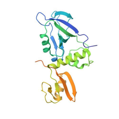
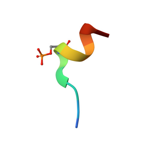
Phosphorylation-dependent recognition of diverse protein targets by the cryptic GK domain of MAGI MAGUKs.

Zhang, M.Cao, A.Lin, L.Chen, Y.Shang, Y.Wang, C.Zhang, M.Zhu, J.

(2023) Sci Adv 9: eadf3295-eadf3295

  • DOI: https://doi.org/10.1126/sciadv.adf3295
  • Primary Citation of Related Structures:  
    7YKF, 7YKG, 7YKH, 7YKI

  • PubMed Abstract: 

    Dynamic signal transduction requires the rapid assembly and disassembly of signaling complexes, often mediated by phosphoprotein binding modules. The guanylate kinase-like (GK) domain of the membrane-associated guanylate kinases (MAGUKs) is such a module orchestrating signaling at cellular junctions. The MAGI subfamily of MAGUKs contains a truncated GK domain with unknown structure and function, although they participate in diverse physiological and pathological processes. Here, we demonstrate that the truncated GK domain of MAGI2 interacts with its adjacent PDZ0 domain to form a structural supramodule capable of recognizing phosphoproteins. A conserved phosphorylation-dependent binding motif for PDZ0-GK is delineated, which leads to identification of a set of previously unknown binding partners. We explore the structure and function of the MAGI2-target complex with an inhibitory peptide derived from the consensus motif. Our work reveals an action mechanism of the cryptic MAGI GKs and broadens our understanding of the target recognition rules of phosphoprotein binding modules.


  • Organizational Affiliation

    Bio-X Institutes, Key Laboratory for the Genetics of Developmental and Neuropsychiatric Disorders, Ministry of Education, Shanghai Jiao Tong University, Shanghai 200240, China.


Macromolecules
Find similar proteins by:  (by identity cutoff)  |  3D Structure
Entity ID: 1
MoleculeChains Sequence LengthOrganismDetailsImage
Membrane-associated guanylate kinase, WW and PDZ domain-containing protein 2
A, C
234Mus musculusMutation(s): 0 
UniProt & NIH Common Fund Data Resources
Find proteins for Q9WVQ1 (Mus musculus)
Explore Q9WVQ1 
Go to UniProtKB:  Q9WVQ1
IMPC:  MGI:1354953
Entity Groups  
Sequence Clusters30% Identity50% Identity70% Identity90% Identity95% Identity100% Identity
UniProt GroupQ9WVQ1
Sequence Annotations
Expand
  • Reference Sequence

Find similar proteins by:  Sequence   |   3D Structure  

Entity ID: 2
MoleculeChains Sequence LengthOrganismDetailsImage
Ephexin4
B, D
12Mus musculusMutation(s): 0 
UniProt & NIH Common Fund Data Resources
Find proteins for Q3U5C8 (Mus musculus)
Explore Q3U5C8 
Go to UniProtKB:  Q3U5C8
IMPC:  MGI:2446219
Entity Groups  
Sequence Clusters30% Identity50% Identity70% Identity90% Identity95% Identity100% Identity
UniProt GroupQ3U5C8
Sequence Annotations
Expand
  • Reference Sequence
Small Molecules
Modified Residues  1 Unique
IDChains TypeFormula2D DiagramParent
SEP
Query on SEP
B, D
L-PEPTIDE LINKINGC3 H8 N O6 PSER
Experimental Data & Validation

Experimental Data

  • Method: X-RAY DIFFRACTION
  • Resolution: 2.28 Å
  • R-Value Free: 0.236 
  • R-Value Work: 0.186 
  • R-Value Observed: 0.188 
  • Space Group: P 1
Unit Cell:
Length ( Å )Angle ( ˚ )
a = 40.209α = 65.31
b = 55.616β = 76.62
c = 61.07γ = 71.56
Software Package:
Software NamePurpose
HKL-2000data scaling
PHENIXrefinement
PDB_EXTRACTdata extraction
HKL-2000data reduction
PHASERphasing

Structure Validation

View Full Validation Report


Entry History & Funding Information

Deposition Data


Funding OrganizationLocationGrant Number
Not funded--

Revision History  (Full details and data files)

  • Version 1.0: 2023-08-02
    Type: Initial release
  • Version 1.1: 2024-02-28
    Changes: Data collection, Database references