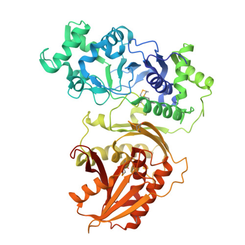
Specific recognition of cyclic oligonucleotides by Cap4 for phage infection.
Chang, J.J., You, B.J., Tien, N., Wang, Y.C., Yang, C.S., Hou, M.H., Chen, Y.(2023) Int J Biol Macromol 237: 123656-123656
- PubMed: 36796558 Search on PubMed
- DOI: https://doi.org/10.1016/j.ijbiomac.2023.123656
- Primary Citation Related Structures:
7YIA, 7YIB - PubMed Abstract:
Under selective pressure, bacteria have evolved diverse defense systems against phage infections. The SMODS-associated and fused to various effector domains (SAVED)-domain containing proteins were identified as major downstream effectors in cyclic oligonucleotide-based antiphage signaling system (CBASS) for bacterial defense. Recent study structurally characterizes a cGAS/DncV-like nucleotidyltransferase (CD-NTase)-associated protein 4 from Acinetobacter baumannii (AbCap4) in complex with 2'3'3'-cyclic AMP-AMP-AMP (cAAA). However, the homologue Cap4 from Enterobacter cloacae (EcCap4) is activated by 3'3'3'-cyclic AMP-AMP-GMP (cAAG). To elucidate the ligand specificity of Cap4 proteins, we determined the crystal structures of full-length wild-type and K74A mutant of EcCap4 to 2.18 and 2.42 Å resolution, respectively. The DNA endonuclease domain of EcCap4 shares similar catalytic mechanism with type II restriction endonuclease. Mutating the key residue K74 in the conserved DX n (D/E)XK motif completely abolishes its DNA degradation activity. The potential ligand-binding cavity of EcCap4 SAVED domain is located adjacent to its N-terminal domain, significantly differing from the centrally located cavity of AbCap4 SAVED domain which recognizes cAAA. Based on structural and bioinformatic analysis, we found that Cap4 proteins can be classified into two types: the type I Cap4, like AbCap4, recognize cAAA and the type II Cap4, like EcCap4, bind cAAG. Several conserved residues identified at the surface of potential ligand-binding pocket of EcCap4 SAVED domain are confirmed by ITC experiment for their direct binding roles for cAAG. Changing Q351, T391 and R392 to alanine abolished the binding of cAAG by EcCap4 and significantly reduced the anti-phage ability of the E. cloacae CBASS system constituting EcCdnD (CD-NTase in clade D) and EcCap4. In summary, we revealed the molecular basis for specific cAAG recognition by the C-terminal SAVED domain of EcCap4 and demonstrates the structural differences for ligand discrimination among different SAVED-domain containing proteins.
- Graduate Institute of Integrated Medicine, China Medical University, Taichung 406, Taiwan; Department of Medical Research, China Medical University Hospital, Taichung 406, Taiwan.
Organizational Affiliation: