7Y3I

Structure of DNA bound SALL4


Experimental Data Snapshot

  • Method: X-RAY DIFFRACTION
  • Resolution: 2.45 Å
  • R-Value Free: 0.242 
  • R-Value Work: 0.194 
  • R-Value Observed: 0.197 

wwPDB Validation   3D Report Full Report


This is version 1.2 of the entry. See complete history


Literature

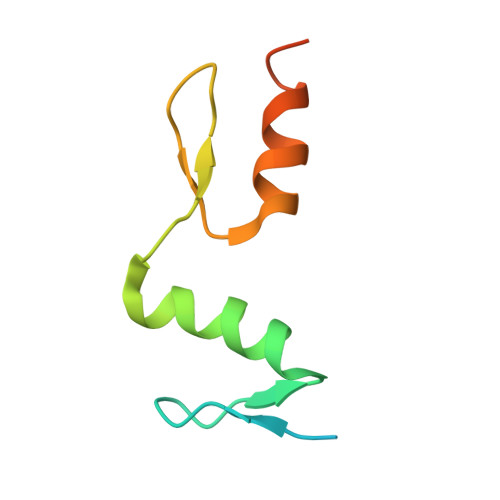
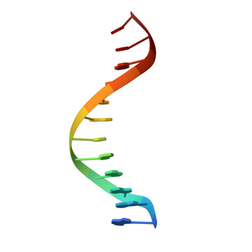
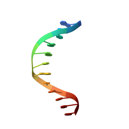
Structural studies of SALL family protein zinc finger cluster domains in complex with DNA reveal preferential binding to an AATA tetranucleotide motif.

Ru, W.Koga, T.Wang, X.Guo, Q.Gearhart, M.D.Zhao, S.Murphy, M.Kawakami, H.Corcoran, D.Zhang, J.Zhu, Z.Yao, X.Kawakami, Y.Xu, C.

(2022) J Biol Chem 298: 102607-102607

  • DOI: https://doi.org/10.1016/j.jbc.2022.102607
  • Primary Citation of Related Structures:  
    7Y3I, 7Y3K, 7Y3L, 7Y3M

  • PubMed Abstract: 

    The Spalt-like 4 transcription factor (SALL4) plays an essential role in controlling the pluripotent property of embryonic stem cells via binding to AT-rich regions of genomic DNA, but structural details on this binding interaction have not been fully characterized. Here, we present crystal structures of the zinc finger cluster 4 (ZFC4) domain of SALL4 (SALL4 ZFC4 ) bound with different dsDNAs containing a conserved AT-rich motif. In the structures, two zinc fingers of SALL4 ZFC4 recognize an AATA tetranucleotide. We also solved the DNA-bound structures of SALL3 ZFC4 and SALL4 ZFC1 . These structures illuminate a common preference for the AATA tetranucleotide shared by ZFC4 of SALL1, SALL3, and SALL4. Furthermore, our cell biology experiments demonstrate that the DNA-binding activity is essential for SALL4 function as DNA-binding defective mutants of mouse Sall4 failed to repress aberrant gene expression in Sall4-/- mESCs. Thus, these analyses provide new insights into the mechanisms of action underlying SALL family proteins in controlling cell fate via preferential targeting to AT-rich sites within genomic DNA during cell differentiation.


  • Organizational Affiliation

    MOE Key Laboratory for Cellular Dynamics, Hefei National Center for Cross-disciplinary Sciences, School of Life Sciences, Division of Life Sciences and Medicine, University of Science and Technology of China, Hefei, P. R. China.


Macromolecules

Find similar proteins by:  (by identity cutoff)  |  3D Structure
Entity ID: 3
MoleculeChains Sequence LengthOrganismDetailsImage
Sal-like protein 4E [auth A],
F [auth B],
G [auth C],
H [auth D]
78Homo sapiensMutation(s): 0 
Gene Names: SALL4ZNF797
UniProt & NIH Common Fund Data Resources
Find proteins for Q9UJQ4 (Homo sapiens)
Explore Q9UJQ4 
Go to UniProtKB:  Q9UJQ4
PHAROS:  Q9UJQ4
GTEx:  ENSG00000101115 
Entity Groups  
Sequence Clusters30% Identity50% Identity70% Identity90% Identity95% Identity100% Identity
UniProt GroupQ9UJQ4
Sequence Annotations
Expand
  • Reference Sequence

Find similar nucleic acids by:  Sequence   |   3D Structure  

Entity ID: 1
MoleculeChains LengthOrganismImage
DNA (12-mer)A [auth L],
C [auth G]
12synthetic construct
Sequence Annotations
Expand
  • Reference Sequence

Find similar nucleic acids by:  Sequence   |   3D Structure  

Entity ID: 2
MoleculeChains LengthOrganismImage
DNA (12-mer)B [auth K],
D [auth H]
12synthetic construct
Sequence Annotations
Expand
  • Reference Sequence
Experimental Data & Validation

Experimental Data

  • Method: X-RAY DIFFRACTION
  • Resolution: 2.45 Å
  • R-Value Free: 0.242 
  • R-Value Work: 0.194 
  • R-Value Observed: 0.197 
  • Space Group: P 21 21 21
Unit Cell:
Length ( Å )Angle ( ˚ )
a = 48.975α = 90
b = 69.197β = 90
c = 110.295γ = 90
Software Package:
Software NamePurpose
PHENIXrefinement
PDB_EXTRACTdata extraction
HKL-2000data reduction
HKL-2000data scaling
PHASERphasing

Structure Validation

View Full Validation Report


Entry History & Funding Information

Deposition Data

  • Released Date: 2022-10-26 
  • Deposition Author(s): Ru, W., Xu, C.

Funding OrganizationLocationGrant Number
National Natural Science Foundation of China (NSFC)China--

Revision History  (Full details and data files)

  • Version 1.0: 2022-10-26
    Type: Initial release
  • Version 1.1: 2022-11-02
    Changes: Database references
  • Version 1.2: 2022-11-30
    Changes: Database references