Structural analysis of the siderophore-interacting protein from Vibrio anguillarum and its implications in classification of Vibrio homologs.
Liu, C., Han, Y., Ma, Q.(2024) Biochem Biophys Res Commun 739: 150979-150979
- PubMed: 39549339 Search on PubMed
- DOI: https://doi.org/10.1016/j.bbrc.2024.150979
- Primary Citation Related Structures:
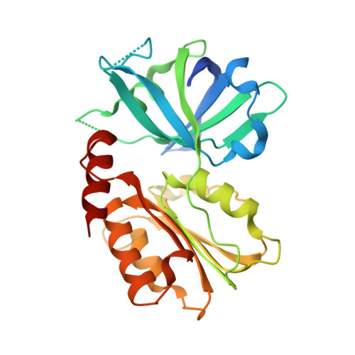
7XLZ - PubMed Abstract:
Bacteria secrete siderophores to sequester the scarce iron in the environments, then the iron is transported into the cell in a siderophore-complexed form, which can be released by siderophore-interacting protein (SIP). Vibrio species comprise an array of serious pathogens, whose iron releasing process by SIP remains poorly understood. Herein, we report the high-resolution (1.2 Å) structure of Vibrio anguillarum SIP (VaSIP) in complex with FAD, representing the first structure of Vibrio SIP. VaSIP consists of a FAD-bound β-barrel domain and a Rossmann-fold domain connected by a linker, like other subgroup I SIPs. FAD is bound to the inter-domain cavity by aromatic stacking and hydrogen bonding interactions. Structural comparison indicated a modified NAD(P)H-binding motif (DxTA-EVL-GE) for subgroup I SIPs. The putative siderophore-binding pocket of VaSIP contains three lysines to form the basic triad to bind siderophore. Phylogenetic analysis shows Vibrio SIPs are mainly divided into two clades, represented by VaSIP and Vibrio cholerae ViuB, respectively. Interestingly, the two clades adopt distinct siderophore-binding basic triads, suggesting functional divergence among Vibrio SIPs. Our results shed light on the structural and phylogenetic characteristics of Vibrio SIPs, providing molecular basis for understanding Vibrio iron metabolism and designing anti-Vibrio drugs.
- CAS and Shandong Province Key Laboratory of Experimental Marine Biology, Institute of Oceanology, Chinese Academy of Sciences, Qingdao, China; Laboratory for Marine Biology and Biotechnology, Qingdao Marine Science and Technology Center, Qingdao, China.
Organizational Affiliation: