7XLY

Crystal structure of FadA2 (Rv0243) from the fatty acid metabolic pathway of Mycobacterium tuberculosis


Experimental Data Snapshot

  • Method: X-RAY DIFFRACTION
  • Resolution: 2.90 Å
  • R-Value Free: 0.285 
  • R-Value Work: 0.245 
  • R-Value Observed: 0.247 

wwPDB Validation   3D Report Full Report


This is version 1.1 of the entry. See complete history


Literature

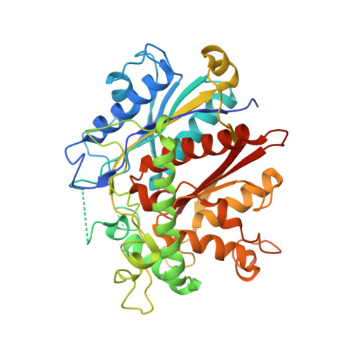
Crystal structure of FadA2 thiolase from Mycobacterium tuberculosis and prediction of its substrate specificity and membrane-anchoring properties.

Singh, R.Kundu, P.Mishra, V.K.Singh, B.K.Bhattacharyya, S.Das, A.K.

(2023) FEBS J 290: 3997-4022

  • DOI: https://doi.org/10.1111/febs.16792
  • Primary Citation of Related Structures:  
    7XLY

  • PubMed Abstract: 

    Tuberculosis (TB) is one of the leading causes of human death caused by Mycobacterium tuberculosis (Mtb). Mtb can enter into a long-lasting persistence where it can utilize fatty acids as the carbon source. Hence, fatty acid metabolism pathway enzymes are considered promising and pertinent mycobacterial drug targets. FadA2 (thiolase) is one of the enzymes involved in Mtb's fatty acid metabolism pathway. FadA2 deletion construct (ΔL136-S150) was designed to produce soluble protein. The crystal structure of FadA2 (ΔL136-S150) at 2.9 Å resolution was solved and analysed for membrane-anchoring region. The four catalytic residues of FadA2 are Cys99, His341, His390 and Cys427, and they belong to four loops with characteristic sequence motifs, i.e., CxT, HEAF, GHP and CxA. FadA2 is the only thiolase of Mtb which belongs to the CHH category containing the HEAF motif. Analysing the substrate-binding channel, it has been suggested that FadA2 is involved in the β-oxidation pathway, i.e., the degradative pathway, as the long-chain fatty acid can be accommodated in the channel. The catalysed reaction is favoured by the presence of two oxyanion holes, i.e., OAH1 and OAH2. OAH1 formation is unique in FadA2, formed by the NE2 of His390 present in the GHP motif and NE2 of His341 present in the HEAF motif, whereas OAH2 formation is similar to CNH category thiolase. Sequence and structural comparison with the human trifunctional enzyme (HsTFE-β) suggests the membrane-anchoring region in FadA2. Molecular dynamics simulations of FadA2 with a membrane containing POPE lipid were conducted to understand the role of a long insertion sequence of FadA2 in membrane anchoring.


  • Organizational Affiliation

    Department of Biotechnology, Indian Institute of Technology Kharagpur, India.


Macromolecules
Find similar proteins by:  (by identity cutoff)  |  3D Structure
Entity ID: 1
MoleculeChains Sequence LengthOrganismDetailsImage
Probable acetyl-CoA acyltransferase FadA2 (3-ketoacyl-CoA thiolase) (Beta-ketothiolase)A [auth C],
B [auth A],
C [auth B],
D
440Mycobacterium tuberculosis H37RvMutation(s): 0 
Gene Names: fadA2Rv0243
EC: 2.3.1.9
UniProt
Find proteins for O86361 (Mycobacterium tuberculosis (strain ATCC 25618 / H37Rv))
Explore O86361 
Go to UniProtKB:  O86361
Entity Groups  
Sequence Clusters30% Identity50% Identity70% Identity90% Identity95% Identity100% Identity
UniProt GroupO86361
Sequence Annotations
Expand
  • Reference Sequence
Small Molecules
Ligands 1 Unique
IDChains Name / Formula / InChI Key2D Diagram3D Interactions
SO4
Query on SO4

Download Ideal Coordinates CCD File 
E [auth B]SULFATE ION
O4 S
QAOWNCQODCNURD-UHFFFAOYSA-L
Modified Residues  1 Unique
IDChains TypeFormula2D DiagramParent
CSX
Query on CSX
A [auth C],
B [auth A],
C [auth B],
D
L-PEPTIDE LINKINGC3 H7 N O3 SCYS
Experimental Data & Validation

Experimental Data

  • Method: X-RAY DIFFRACTION
  • Resolution: 2.90 Å
  • R-Value Free: 0.285 
  • R-Value Work: 0.245 
  • R-Value Observed: 0.247 
  • Space Group: P 32 2 1
Unit Cell:
Length ( Å )Angle ( ˚ )
a = 109.782α = 90
b = 109.782β = 90
c = 353.112γ = 120
Software Package:
Software NamePurpose
SCALAdata scaling
PHENIXrefinement
PDB_EXTRACTdata extraction
XDSdata reduction
PHASERphasing

Structure Validation

View Full Validation Report


Entry History & Funding Information

Deposition Data


Funding OrganizationLocationGrant Number
Department of Biotechnology (DBT, India)IndiaBT/PR12404/BRB/10/1362/2014

Revision History  (Full details and data files)

  • Version 1.0: 2023-04-26
    Type: Initial release
  • Version 1.1: 2023-08-30
    Changes: Data collection, Database references