7XLL | pdb_00007xll

Alanine racemase from Lactobacillus sakei Uonuma-1.


Experimental Data Snapshot

  • Method: X-RAY DIFFRACTION
  • Resolution: 1.76 Å
  • R-Value Free: 
    0.199 (Depositor), 0.200 (DCC) 
  • R-Value Work: 
    0.165 (Depositor), 0.166 (DCC) 
  • R-Value Observed: 
    0.166 (Depositor) 

Starting Model: experimental
View more details

wwPDB Validation 3D Report Full Report

Validation slider image for 7XLL

This is version 1.2 of the entry. See complete history

Literature

Regulation of alanine racemase activity by carboxylates and the d-type substrate d-alanine.

Shimizu-Ibuka, A.Sato, A.Ichimura, H.Hiraga, H.Nakayama, S.Nishiwaki, T.

(2023) FEBS J 290: 2954-2967

  • DOI: https://doi.org/10.1111/febs.16745
  • Primary Citation Related Structures: 
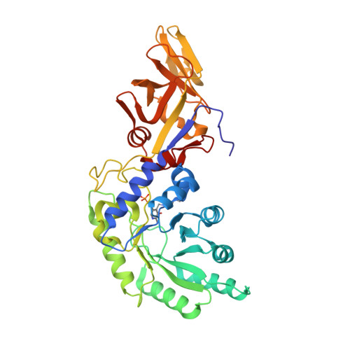
    7XLL

  • PubMed Abstract: 

    Alanine racemases (ALRs) are essential for d-alanine (d-Ala) production in bacteria, and many ALRs have a conserved carbamylated lysine residue in the active site. Although short-chain carboxylates inhibit ALRs harbouring this lysine residue as substrate analogues, in an ALR variant with an alanine residue at this position, carboxylates behave as activators; however, this activation mechanism remains unclear. Here, we performed kinetic and structural analyses of U1ALR, an ALR from Latilactobacillus sakei UONUMA harbouring a glycine residue (Gly134) in the site of the carbamylated lysine residue. U1ALR was activated by various carboxylates and also by a G134K mutation, both of which caused a significant decrease in K m , indicating an increase in substrate affinity. The U1ALR crystal structure revealed the presence of an acetate molecule bound in a position and at an orientation resembling the conformation of the carbamylated lysine side chain observed in the structures of other ALRs. These results suggest a regulatory mechanism for U1ALR activity involving two carboxylate-binding sites: one with high affinity near Gly134, where an acetate molecule is observed in the crystal structure and carboxylate binding results in enzyme activation; the other is the substrate-binding site, where carboxylate binding inhibits enzyme activity. Furthermore, we observed no carboxylate/G134K-mediated activation in the presence of d-Ala at high concentrations, implying that d-Ala also exhibits low-affinity binding in the first carboxylate-binding site and prevents carboxylate/G134K-induced activation. Such regulation of enzyme activity by carboxylates and d-Ala may be ubiquitous in many ALRs from lactic acid bacteria sharing the same sequence characteristics.


  • Organizational Affiliation
    • Department of Applied Life Sciences, Niigata University of Pharmacy and Applied Life Sciences, Japan.

Macromolecule Content 

  • Total Structure Weight: 84.45 kDa 
  • Atom Count: 6,856 
  • Modeled Residue Count: 756 
  • Deposited Residue Count: 760 
  • Unique protein chains: 1

Macromolecules

Find similar proteins by:|  3D Structure
Entity ID: 1
MoleculeChains  Sequence LengthOrganismDetailsImage
Alanine racemase
A, B
380Latilactobacillus sakeiMutation(s): 0 
Gene Names: C0213_08900
EC: 5.1.1.1

Small Molecules

Ligands 3 Unique
IDChains Name / Formula / InChI Key2D Diagram3D Interactions
PEG

Query on PEG


Download:Ideal Coordinates CCD File
F [auth A]DI(HYDROXYETHYL)ETHER
C4 H10 O3
MTHSVFCYNBDYFN-UHFFFAOYSA-N
ALA

Query on ALA


Download:Ideal Coordinates CCD File
G [auth A]ALANINE
C3 H7 N O2
QNAYBMKLOCPYGJ-REOHCLBHSA-N
ACT

Query on ACT


Download:Ideal Coordinates CCD File
C [auth A]
D [auth A]
E [auth A]
H [auth B]
I [auth B]
C [auth A],
D [auth A],
E [auth A],
H [auth B],
I [auth B],
J [auth B]
ACETATE ION
C2 H3 O2
QTBSBXVTEAMEQO-UHFFFAOYSA-M
Modified Residues  1 Unique
IDChains TypeFormula2D DiagramParent
LLP
Query on LLP
A, B
L-PEPTIDE LINKINGC14 H22 N3 O7 PLYS

Experimental Data & Validation

Experimental Data

  • Method: X-RAY DIFFRACTION
  • Resolution: 1.76 Å
  • R-Value Free:  0.199 (Depositor), 0.200 (DCC) 
  • R-Value Work:  0.165 (Depositor), 0.166 (DCC) 
  • R-Value Observed: 0.166 (Depositor) 
Space Group: P 21 21 2
Unit Cell:
Length ( Å )Angle ( ˚ )
a = 107.73α = 90
b = 125.04β = 90
c = 56.64γ = 90
Software Package:
Software NamePurpose
PHENIXrefinement
MOSFLMdata reduction
SCALAdata scaling
MOLREPphasing

Structure Validation

View Full Validation Report


Entry History 

& Funding Information

Deposition Data


Funding OrganizationLocationGrant Number
Not funded--

Revision History  (Full details and data files)

  • Version 1.0: 2023-03-01
    Type: Initial release
  • Version 1.1: 2023-06-21
    Changes: Database references
  • Version 1.2: 2023-11-29
    Changes: Data collection, Refinement description