7WR1

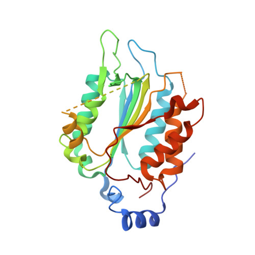
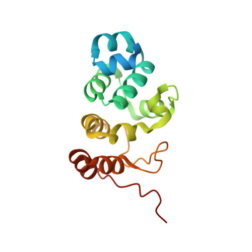
P32 of caspase-4 C258A mutant in complex with OspC3 C-terminal ankyrin-repeat domain


Experimental Data Snapshot

  • Method: X-RAY DIFFRACTION
  • Resolution: 2.13 Å
  • R-Value Free: 0.200 
  • R-Value Work: 0.170 
  • R-Value Observed: 0.171 

wwPDB Validation   3D Report Full Report


This is version 1.3 of the entry. See complete history


Literature

Structural mechanisms of calmodulin activation of Shigella effector OspC3 to ADP-riboxanate caspase-4/11 and block pyroptosis.

Hou, Y.Zeng, H.Li, Z.Feng, N.Meng, F.Xu, Y.Li, L.Shao, F.Ding, J.

(2023) Nat Struct Mol Biol 30: 261-272

  • DOI: https://doi.org/10.1038/s41594-022-00888-3
  • Primary Citation of Related Structures:  
    7WR0, 7WR1, 7WR2, 7WR3, 7WR4, 7WR5, 7WR6

  • PubMed Abstract: 

    The caspase-4/11-GSDMD pyroptosis axis recognizes cytosolic lipopolysaccharide for antibacterial defenses. Shigella flexneri employs an OspC3 effector to block pyroptosis by catalyzing NAD + -dependent arginine ADP-riboxanation of caspase-4/11. Here, we identify Ca 2+ -free calmodulin (CaM) that binds and stimulates OspC3 ADP-riboxanase activity. Crystal structures of OspC3-CaM and OspC3-caspase-4 binary complexes reveal unique CaM binding to an OspC3 N-terminal domain featuring an ADP-ribosyltransferase-like fold and specific recognition of caspase-4 by an OspC3 ankryin repeat domain, respectively. CaM-OspC3-caspase-4 ternary complex structures show that NAD + binding reorganizes the catalytic pocket, in which D231 and D177 activate the substrate arginine for initial ADP-ribosylation and ribosyl 2'-OH in the ADP-ribosylated arginine, respectively, for subsequent deamination. We also determine structures of unmodified and OspC3-ADP-riboxanated caspase-4. Mechanisms derived from this series of structures covering the entire process of OspC3 action are supported by biochemical analyses in vitro and functional validation in S. flexneri-infected mice.


  • Organizational Affiliation

    National Laboratory of Biomacromolecules, CAS Center for Excellence in Biomacromolecules, Institute of Biophysics, Chinese Academy of Sciences, Beijing, China.


Macromolecules
Find similar proteins by:  (by identity cutoff)  |  3D Structure
Entity ID: 1
MoleculeChains Sequence LengthOrganismDetailsImage
Caspase-4
A, B
280Homo sapiensMutation(s): 1 
Gene Names: CASP4
EC: 3.4.22.57
UniProt & NIH Common Fund Data Resources
Find proteins for P49662 (Homo sapiens)
Explore P49662 
Go to UniProtKB:  P49662
PHAROS:  P49662
GTEx:  ENSG00000196954 
Entity Groups  
Sequence Clusters30% Identity50% Identity70% Identity90% Identity95% Identity100% Identity
UniProt GroupP49662
Sequence Annotations
Expand
  • Reference Sequence
Find similar proteins by:  (by identity cutoff)  |  3D Structure
Entity ID: 2
MoleculeChains Sequence LengthOrganismDetailsImage
OspC3
C, D
161Shigella flexneriMutation(s): 0 
Gene Names: ospC3
UniProt
Find proteins for A0A0H2US87 (Shigella flexneri)
Explore A0A0H2US87 
Go to UniProtKB:  A0A0H2US87
Entity Groups  
Sequence Clusters30% Identity50% Identity70% Identity90% Identity95% Identity100% Identity
UniProt GroupA0A0H2US87
Sequence Annotations
Expand
  • Reference Sequence
Experimental Data & Validation

Experimental Data

  • Method: X-RAY DIFFRACTION
  • Resolution: 2.13 Å
  • R-Value Free: 0.200 
  • R-Value Work: 0.170 
  • R-Value Observed: 0.171 
  • Space Group: P 1 21 1
Unit Cell:
Length ( Å )Angle ( ˚ )
a = 76.639α = 90
b = 59.751β = 104.083
c = 121.746γ = 90
Software Package:
Software NamePurpose
PHENIXrefinement
XDSdata reduction
XDSdata scaling
PHASERphasing

Structure Validation

View Full Validation Report


Entry History & Funding Information

Deposition Data


Funding OrganizationLocationGrant Number
Chinese Academy of SciencesChina--

Revision History  (Full details and data files)

  • Version 1.0: 2023-01-25
    Type: Initial release
  • Version 1.1: 2023-02-01
    Changes: Database references
  • Version 1.2: 2023-03-29
    Changes: Database references
  • Version 1.3: 2023-11-29
    Changes: Data collection, Refinement description