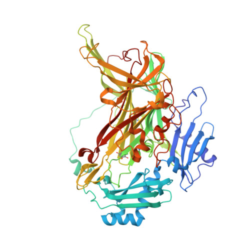
Re-evaluation of protein neutron crystallography with and without X-ray/neutron joint refinement.
Murakawa, T., Kurihara, K., Adachi, M., Kusaka, K., Tanizawa, K., Okajima, T.(2022) IUCrJ 9: 342-348
- PubMed: 35546796
- DOI: https://doi.org/10.1107/S2052252522003657
- Primary Citation of Related Structures:
7WNO, 7WNP - PubMed Abstract:
Protein neutron crystallography is a powerful technique to determine the positions of H atoms, providing crucial biochemical information such as the protonation states of catalytic groups and the geometry of hydrogen bonds. Recently, the crystal structure of a bacterial copper amine oxidase was determined by joint refinement using X-ray and neutron diffraction data sets at resolutions of 1.14 and 1.72 Å, respectively [Murakawa et al. (2020 ▸). Proc. Natl Acad. Sci. USA , 117 , 10818-10824]. While joint refinement is effective for the determination of the accurate positions of heavy atoms on the basis of the electron density, the structural information on light atoms (hydrogen and deuterium) derived from the neutron diffraction data might be affected by the X-ray data. To unravel the information included in the neutron diffraction data, the structure determination was conducted again using only the neutron diffraction data at 1.72 Å resolution and the results were compared with those obtained in the previous study. Most H and D atoms were identified at essentially the same positions in both the neutron-only and the X-ray/neutron joint refinements. Nevertheless, neutron-only refinement was found to be less effective than joint refinement in providing very accurate heavy-atom coordinates that lead to significant improvement of the neutron scattering length density map, especially for the active-site cofactor. Consequently, it was confirmed that X-ray/neutron joint refinement is crucial for determination of the real chemical structure of the catalytic site of the enzyme.
- Department of Biochemistry, Faculty of Medicine, Osaka Medical and Pharmaceutical University, 2-7 Daigakumachi, Takatsuki, Osaka 569-8686, Japan.
Organizational Affiliation:


