A Structural and Bioinformatics Investigation of a Fungal Squalene Synthase and Comparisons with Other Membrane Proteins.
Malwal, S.R., Shang, N., Liu, W., Li, X., Zhang, L., Chen, C.C., Guo, R.T., Oldfield, E.(2022) ACS Omega 7: 22601-22612
- PubMed: 35811857 Search on PubMedSearch on PubMed Central
- DOI: https://doi.org/10.1021/acsomega.2c01924
- Primary Citation Related Structures:
7WGH, 7WGI - PubMed Abstract:
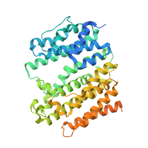
There is interest in the development of drugs to treat fungal infections due to the increasing threat of drug resistance, and here, we report the first crystallographic structure of the catalytic domain of a fungal squalene synthase (SQS), Aspergillus flavus SQS (AfSQS), a potential drug target, together with a bioinformatics study of fungal, human, and protozoal SQSs. Our X-ray results show strong structural similarities between the catalytic domains in these proteins, but, remarkably, using bioinformatics, we find that there is also a large, highly polar helix in the fungal proteins that connects the catalytic and membrane-anchoring transmembrane domains. This polar helix is absent in squalene synthases from all other lifeforms. We show that the transmembrane domain in AfSQS and in other SQSs, stannin, and steryl sulfatase, have very similar properties (% polar residues, hydrophobicity, and hydrophobic moment) to those found in the "penultimate" C-terminal helical domain in squalene epoxidase, while the final C-terminal domain in squalene epoxidase is more polar and may be monotopic. We also propose structural models for full-length AfSQS based on the bioinformatics results as well as a deep learning program that indicate that the C-terminus region may also be membrane surface-associated. Taken together, our results are of general interest given the unique nature of the polar helical domain in fungi that may be involved in protein-protein interactions as well as being a future target for antifungals.
- Department of Chemistry, University of Illinois at Urbana-Champaign, Urbana, Illinois 61801, United States.
Organizational Affiliation: