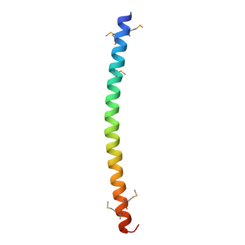
7W91

Residues 440-490 of centrosomal protein 63


Experimental Data Snapshot

  • Method: X-RAY DIFFRACTION
  • Resolution: 3.29 Å
  • R-Value Free: 0.261 
  • R-Value Work: 0.229 
  • R-Value Observed: 0.232 

wwPDB Validation   3D Report Full Report


This is version 1.0 of the entry. See complete history


Literature

Architectural basis for cylindrical self-assembly governing Plk4-mediated centriole duplication in human cells.

Il Ahn, J.Zhang, L.Ravishankar, H.Fan, L.Kirsch, K.Zeng, Y.Meng, L.Park, J.E.Yun, H.Y.Ghirlando, R.Ma, B.Ball, D.Ku, B.Nussinov, R.Schmit, J.D.Heinz, W.F.Kim, S.J.Karpova, T.Wang, Y.X.Lee, K.S.

(2023) Commun Biol 6: 712-712

  • DOI: https://doi.org/10.1038/s42003-023-05067-8
  • Primary Citation of Related Structures:  
    7W91

  • PubMed Abstract: 

    Proper organization of intracellular assemblies is fundamental for efficient promotion of biochemical processes and optimal assembly functionality. Although advances in imaging technologies have shed light on how the centrosome is organized, how its constituent proteins are coherently architected to elicit downstream events remains poorly understood. Using multidisciplinary approaches, we showed that two long coiled-coil proteins, Cep63 and Cep152, form a heterotetrameric building block that undergoes a stepwise formation into higher molecular weight complexes, ultimately generating a cylindrical architecture around a centriole. Mutants defective in Cep63•Cep152 heterotetramer formation displayed crippled pericentriolar Cep152 organization, polo-like kinase 4 (Plk4) relocalization to the procentriole assembly site, and Plk4-mediated centriole duplication. Given that the organization of pericentriolar materials (PCM) is evolutionarily conserved, this work could serve as a model for investigating the structure and function of PCM in other species, while offering a new direction in probing the organizational defects of PCM-related human diseases.


  • Organizational Affiliation

    Cancer Innovation Laboratory, Center for Cancer Research, National Cancer Institute, National Institutes of Health, Bethesda, MD, 20892, USA.


Macromolecules
Find similar proteins by:  (by identity cutoff)  |  3D Structure
Entity ID: 1
MoleculeChains Sequence LengthOrganismDetailsImage
Centrosomal protein of 63 kDa
A, B, C, D, E
A, B, C, D, E, F, G, H, I, J, K, L
56Homo sapiensMutation(s): 0 
Gene Names: CEP63
UniProt & NIH Common Fund Data Resources
Find proteins for Q96MT8 (Homo sapiens)
Explore Q96MT8 
Go to UniProtKB:  Q96MT8
PHAROS:  Q96MT8
GTEx:  ENSG00000182923 
Entity Groups  
Sequence Clusters30% Identity50% Identity70% Identity90% Identity95% Identity100% Identity
UniProt GroupQ96MT8
Sequence Annotations
Expand
  • Reference Sequence
Small Molecules
Modified Residues  1 Unique
IDChains TypeFormula2D DiagramParent
MSE
Query on MSE
A, B, C, D, E
A, B, C, D, E, F, G, H, I, J, K, L
L-PEPTIDE LINKINGC5 H11 N O2 SeMET
Experimental Data & Validation

Experimental Data

  • Method: X-RAY DIFFRACTION
  • Resolution: 3.29 Å
  • R-Value Free: 0.261 
  • R-Value Work: 0.229 
  • R-Value Observed: 0.232 
  • Space Group: P 1
Unit Cell:
Length ( Å )Angle ( ˚ )
a = 80.622α = 65.33
b = 81.112β = 60.07
c = 80.846γ = 60.2
Software Package:
Software NamePurpose
PHENIXrefinement
PDB_EXTRACTdata extraction
HKL-2000data reduction
HKL-2000data scaling
PHENIXphasing

Structure Validation

View Full Validation Report


Entry History & Funding Information

Deposition Data


Funding OrganizationLocationGrant Number
National Research Foundation (NRF, Korea)Korea, Republic Of2019M3E5D6063955
National Research Foundation (NRF, Korea)Korea, Republic OfKGM9952112

Revision History  (Full details and data files)

  • Version 1.0: 2023-07-19
    Type: Initial release