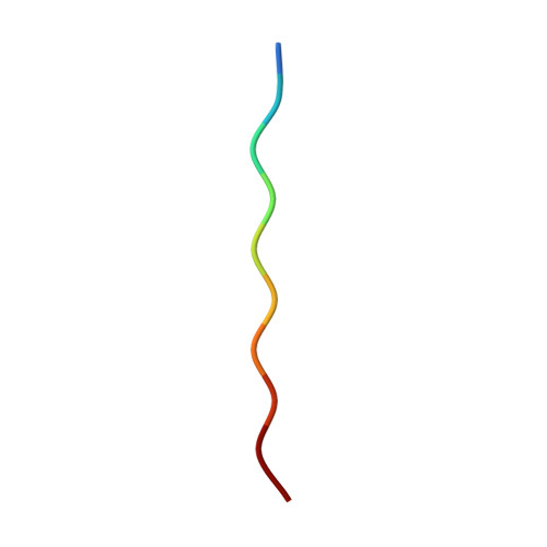
ALS mutations in the TIA-1 prion-like domain trigger highly condensed pathogenic structures.
Sekiyama, N., Takaba, K., Maki-Yonekura, S., Akagi, K.I., Ohtani, Y., Imamura, K., Terakawa, T., Yamashita, K., Inaoka, D., Yonekura, K., Kodama, T.S., Tochio, H.(2022) Proc Natl Acad Sci U S A 119: e2122523119-e2122523119
- PubMed: 36112647
- DOI: https://doi.org/10.1073/pnas.2122523119
- Primary Citation of Related Structures:
7VI4, 7VI5 - PubMed Abstract:
T cell intracellular antigen-1 (TIA-1) plays a central role in stress granule (SG) formation by self-assembly via the prion-like domain (PLD). In the TIA-1 PLD, amino acid mutations associated with neurodegenerative diseases, such as amyotrophic lateral sclerosis (ALS) or Welander distal myopathy (WDM), have been identified. However, how these mutations affect PLD self-assembly properties has remained elusive. In this study, we uncovered the implicit pathogenic structures caused by the mutations. NMR analysis indicated that the dynamic structures of the PLD are synergistically determined by the physicochemical properties of amino acids in units of five residues. Molecular dynamics simulations and three-dimensional electron crystallography, together with biochemical assays, revealed that the WDM mutation E384K attenuated the sticky properties, whereas the ALS mutations P362L and A381T enhanced the self-assembly by inducing β-sheet interactions and highly condensed assembly, respectively. These results suggest that the P362L and A381T mutations increase the likelihood of irreversible amyloid fibrillization after phase-separated droplet formation, and this process may lead to pathogenicity.
- Department of Biophysics, Graduate School of Science, Kyoto University, Kyoto 606-8502, Japan.
Organizational Affiliation:


