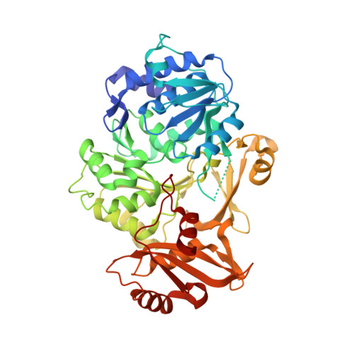
Structural and functional analysis of the D-alanyl carrier protein ligase DltA from Staphylococcus aureus Mu50.
Lee, I.G., Song, C., Yang, S., Jeon, H., Park, J., Yoon, H.J., Im, H., Kang, S.M., Eun, H.J., Lee, B.J.(2022) Acta Crystallogr D Struct Biol 78: 424-434
- PubMed: 35362466 Search on PubMedSearch on PubMed Central
- DOI: https://doi.org/10.1107/S2059798322000547
- Primary Citation Related Structures:
7VHV - PubMed Abstract:
D-Alanylation of the teichoic acids of the Gram-positive bacterial cell wall plays crucial roles in bacterial physiology and virulence. Deprivation of D-alanine from the teichoic acids of Staphylococcus aureus impairs biofilm and colony formation, induces autolysis and ultimately renders methicillin-resistant S. aureus highly susceptible to antimicrobial agents and host defense peptides. Hence, the D-alanylation pathway has emerged as a promising antibacterial target against drug-resistant S. aureus. D-Alanylation of teichoic acids is mediated via the action of four proteins encoded by the dlt operon, DltABCD, all four of which are essential for the process. In order to develop novel antimicrobial agents against S. aureus, the D-alanyl carrier protein ligase DltA, which is the first protein in the D-alanylation pathway, was focused on. Here, the crystal structure of DltA from the methicillin-resistant S. aureus strain Mu50 is presented, which reveals the unique molecular details of the catalytic center and the role of the P-loop. Kinetic analysis shows that the enantioselectivity of S. aureus DltA is much higher than that of DltA from other species. In the presence of DltC, the enzymatic activity of DltA is increased by an order of magnitude, suggesting a new exploitable binding pocket. This discovery may pave the way for a new generation of treatments for drug-resistant S. aureus.
- Chemical Kinomics Research Center, Korea Institute of Science and Technology (KIST), 5 Hwarangro 14-gil, Seongbuk-gu, Seoul 02792, Republic of Korea.
Organizational Affiliation: