Structural Analysis of Hen Egg Lysozyme Refolded after Denaturation at Acidic pH.
Oda, M., Sano, T., Kamatari, Y.O., Abe, Y., Ikura, T., Ito, N.(2022) Protein J 41: 71-78
- PubMed: 35094218 Search on PubMedSearch on PubMed Central
- DOI: https://doi.org/10.1007/s10930-021-10036-3
- Primary Citation Related Structures:
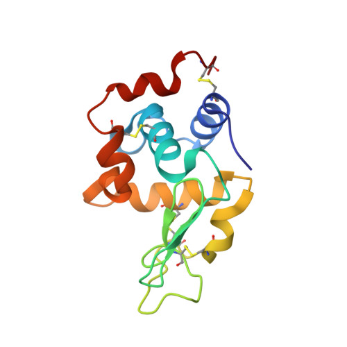
7VGO, 7VGP - PubMed Abstract:
Protein structures fluctuate in solution; therefore, proteins have multiple stable structures that are slightly different from each other. In this study, we determined the crystal structure of hen egg lysozyme refolded after denaturation at acidic pH (rHEL) and found a structure different from native HEL (nHEL). The different local conformations of the peptide bond between Asp101 and Gly102 found in the crystal structure was supported by the NMR results for nHEL and rHEL. The NMR experiments also showed shifts in the heteronuclear single quantum coherence signals derived from Thr43 and Asp52. The chemical shift change of Asp52 could be explained by the crystal structure of rHEL, showing the conformational change of Tyr53, whose phenol ring directly lies on the main chain of Asp52. The catalytic activity of rHEL was similar to that of nHEL, indicating that the conformational change had little effect on activity. In contrast, conformational changes could be detected by the binding of monoclonal antibodies against HEL. Using multiple methods, we successfully detected the unusual structure of HEL, which might be another stable structure of HEL in solution.
- Graduate School of Life and Environmental Sciences, Kyoto Prefectural University, 1-5 Hangi-cho, Shimogamo, Sakyo-ku, Kyoto, 606-8522, Japan. oda@kpu.ac.jp.
Organizational Affiliation: