7VFV | pdb_00007vfv

Human N-type voltage gated calcium channel CaV2.2-alpha2/delta1-beta1 complex, bound to PD173212


Experimental Data Snapshot

  • Method: ELECTRON MICROSCOPY
  • Resolution: 3.00 Å
  • Aggregation State: PARTICLE 
  • Reconstruction Method: SINGLE PARTICLE 

wwPDB Validation 3D Report Full Report

Validation slider image for 7VFV

This is version 1.3 of the entry. See complete history

Literature

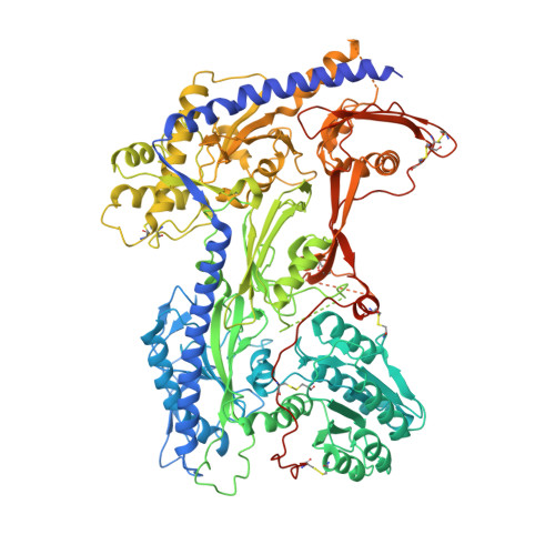
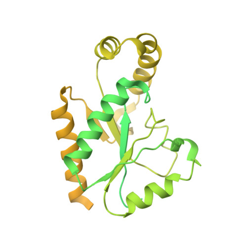
Closed-state inactivation and pore-blocker modulation mechanisms of human Ca V 2.2.

Dong, Y.Gao, Y.Xu, S.Wang, Y.Yu, Z.Li, Y.Li, B.Yuan, T.Yang, B.Zhang, X.C.Jiang, D.Huang, Z.Zhao, Y.

(2021) Cell Rep 37: 109931-109931

  • DOI: https://doi.org/10.1016/j.celrep.2021.109931
  • Primary Citation Related Structures: 
    7VFS, 7VFU, 7VFV, 7VFW

  • PubMed Abstract: 

    N-type voltage-gated calcium (Ca V ) channels mediate Ca 2+ influx at presynaptic terminals in response to action potentials and play vital roles in synaptogenesis, release of neurotransmitters, and nociceptive transmission. Here, we elucidate a cryo-electron microscopy (cryo-EM) structure of the human Ca V 2.2 complex in apo, ziconotide-bound, and two Ca V 2.2-specific pore blockers-bound states. The second voltage-sensing domain (VSD) is captured in a resting-state conformation, trapped by a phosphatidylinositol 4,5-bisphosphate (PIP 2 ) molecule, which is distinct from the other three VSDs of Ca V 2.2, as well as activated VSDs observed in previous structures of Ca V channels. This structure reveals the molecular basis for the unique inactivation process of Ca V 2.2 channels, in which the intracellular gate formed by S6 helices is closed and a W-helix from the domain II-III linker stabilizes closed-state inactivation. The structures of this inactivated, drug-bound complex lay a solid foundation for developing new state-dependent blockers for treatment of chronic pain.


  • Organizational Affiliation
    • National Laboratory of Biomacromolecules, CAS Center for Excellence in Biomacromolecules, Institute of Biophysics, Chinese Academy of Sciences, Beijing 100101, China; State Key Laboratory of Brain and Cognitive Science, Institute of Biophysics, Chinese Academy of Sciences, 15 Datun Road, Beijing 100101, China; College of Life Sciences, University of Chinese Academy of Sciences, Beijing 100049, China.

Macromolecule Content 

  • Total Structure Weight: 463.66 kDa 
  • Atom Count: 19,776 
  • Modeled Residue Count: 2,383 
  • Deposited Residue Count: 4,040 
  • Unique protein chains: 3

Macromolecules

Find similar proteins by:|  3D Structure
Entity ID: 1
MoleculeChains  Sequence LengthOrganismDetailsImage
Voltage-dependent N-type calcium channel subunit alpha-1B2,339Homo sapiensMutation(s): 0 
Gene Names: CACNA1BCACH5CACNL1A5
Membrane Entity: Yes 
UniProt & NIH Common Fund Data Resources
Find proteins for Q00975 (Homo sapiens)
Explore Q00975 
Go to UniProtKB:  Q00975
PHAROS:  Q00975
GTEx:  ENSG00000148408 
Entity Groups
Sequence Clusters30% Identity50% Identity70% Identity90% Identity95% Identity100% Identity
UniProt GroupQ00975
Sequence Annotations
Expand
Reference Sequence
Find similar proteins by:|  3D Structure
Entity ID: 2
MoleculeChains  Sequence LengthOrganismDetailsImage
Voltage-dependent calcium channel subunit alpha-2/delta-11,103Homo sapiensMutation(s): 0 
Gene Names: CACNA2D1CACNL2ACCHL2AMHS3
Membrane Entity: Yes 
UniProt & NIH Common Fund Data Resources
Find proteins for P54289 (Homo sapiens)
Explore P54289 
Go to UniProtKB:  P54289
PHAROS:  P54289
GTEx:  ENSG00000153956 
Entity Groups
Sequence Clusters30% Identity50% Identity70% Identity90% Identity95% Identity100% Identity
UniProt GroupP54289
Glycosylation
Glycosylation Sites: 11Go to GlyGen: P54289-1
Sequence Annotations
Expand
Reference Sequence
Find similar proteins by:|  3D Structure
Entity ID: 3
MoleculeChains  Sequence LengthOrganismDetailsImage
Voltage-dependent L-type calcium channel subunit beta-1C [auth D]598Homo sapiensMutation(s): 0 
Gene Names: CACNB1CACNLB1
Membrane Entity: Yes 
UniProt & NIH Common Fund Data Resources
Find proteins for Q02641 (Homo sapiens)
Explore Q02641 
Go to UniProtKB:  Q02641
PHAROS:  Q02641
GTEx:  ENSG00000067191 
Entity Groups
Sequence Clusters30% Identity50% Identity70% Identity90% Identity95% Identity100% Identity
UniProt GroupQ02641
Sequence Annotations
Expand
Reference Sequence

Oligosaccharides

Help  
Entity ID: 4
MoleculeChains Length2D Diagram GlycosylationD Interactions
beta-D-mannopyranose-(1-4)-2-acetamido-2-deoxy-beta-D-glucopyranose-(1-4)-2-acetamido-2-deoxy-beta-D-glucopyranoseD [auth C],
E
3N-Glycosylation
Glycosylation Resources
GlyTouCan: G15407YE
GlyCosmos: G15407YE
GlyGen: G15407YE
Entity ID: 5
MoleculeChains Length2D Diagram GlycosylationD Interactions
beta-D-mannopyranose-(1-3)-2-acetamido-2-deoxy-beta-D-glucopyranose-(1-4)-2-acetamido-2-deoxy-beta-D-glucopyranose
F
3N-Glycosylation
Glycosylation Resources
GlyTouCan: G44266EX
GlyCosmos: G44266EX
GlyGen: G44266EX

Small Molecules

Ligands 6 Unique
IDChains Name / Formula / InChI Key2D Diagram3D Interactions
PT5
(Subject of Investigation/LOI)

Query on PT5


Download:Ideal Coordinates CCD File
Z [auth A][(2R)-1-octadecanoyloxy-3-[oxidanyl-[(1R,2R,3S,4R,5R,6S)-2,3,6-tris(oxidanyl)-4,5-diphosphonooxy-cyclohexyl]oxy-phospho ryl]oxy-propan-2-yl] (8Z)-icosa-5,8,11,14-tetraenoate
C47 H85 O19 P3
CNWINRVXAYPOMW-HJBQCNPJSA-N
6IX
(Subject of Investigation/LOI)

Query on 6IX


Download:Ideal Coordinates CCD File
AA [auth A](2~{S})-~{N}-[(2~{S})-1-(~{tert}-butylamino)-1-oxidanylidene-3-(4-phenylmethoxyphenyl)propan-2-yl]-2-[(4-~{tert}-butylphenyl)methyl-methyl-amino]-4-methyl-pentanamide
C38 H53 N3 O3
GCDHMGROXQUFNR-HEVIKAOCSA-N
Y01
(Subject of Investigation/LOI)

Query on Y01


Download:Ideal Coordinates CCD File
J [auth A],
P [auth A],
Y [auth A]
CHOLESTEROL HEMISUCCINATE
C31 H50 O4
WLNARFZDISHUGS-MIXBDBMTSA-N
R16
(Subject of Investigation/LOI)

Query on R16


Download:Ideal Coordinates CCD File
G [auth A]
H [auth A]
I [auth A]
K [auth A]
L [auth A]
G [auth A],
H [auth A],
I [auth A],
K [auth A],
L [auth A],
M [auth A],
N [auth A],
O [auth A],
Q [auth A],
R [auth A],
S [auth A],
T [auth A],
U [auth A],
V [auth A],
W [auth A],
X [auth A]
HEXADECANE
C16 H34
DCAYPVUWAIABOU-UHFFFAOYSA-N
NAG

Query on NAG


Download:Ideal Coordinates CCD File
DA [auth B]
EA [auth B]
FA [auth B]
GA [auth B]
HA [auth B]
DA [auth B],
EA [auth B],
FA [auth B],
GA [auth B],
HA [auth B],
IA [auth B],
JA [auth B],
KA [auth B]
2-acetamido-2-deoxy-beta-D-glucopyranose
C8 H15 N O6
OVRNDRQMDRJTHS-FMDGEEDCSA-N
CA

Query on CA


Download:Ideal Coordinates CCD File
BA [auth A],
CA [auth B]
CALCIUM ION
Ca
BHPQYMZQTOCNFJ-UHFFFAOYSA-N
Binding Affinity Annotations 
IDSourceBinding Affinity
6IX BindingDB:  7VFV IC50: 40 (nM) from 1 assay(s)

Experimental Data & Validation

Experimental Data

  • Method: ELECTRON MICROSCOPY
  • Resolution: 3.00 Å
  • Aggregation State: PARTICLE 
  • Reconstruction Method: SINGLE PARTICLE 
EM Software:
TaskSoftware PackageVersion
MODEL REFINEMENTPHENIX

Structure Validation

View Full Validation Report


Entry History 

& Funding Information

Deposition Data


Funding OrganizationLocationGrant Number
Chinese Academy of SciencesChinaXDB37030304

Revision History  (Full details and data files)

  • Version 1.0: 2021-11-03
    Type: Initial release
  • Version 1.1: 2022-02-16
    Changes: Database references
  • Version 1.2: 2025-07-02
    Changes: Data collection, Structure summary
  • Version 1.3: 2025-09-17
    Changes: Advisory, Data collection, Derived calculations, Structure summary