7VEW

Crystal structure of bacterial chemotaxis-dependent pectin-binding protein SPH1118 in complex with unsaturated trigalacturonic acid


Experimental Data Snapshot

  • Method: X-RAY DIFFRACTION
  • Resolution: 1.92 Å
  • R-Value Free: 0.232 
  • R-Value Work: 0.210 

wwPDB Validation   3D Report Full Report


This is version 1.1 of the entry. See complete history


Literature

Substrate size-dependent conformational changes of bacterial pectin-binding protein crucial for chemotaxis and assimilation.

Anamizu, K.Takase, R.Hio, M.Watanabe, D.Mikami, B.Hashimoto, W.

(2022) Sci Rep 12: 12653-12653

  • DOI: https://doi.org/10.1038/s41598-022-16540-5
  • Primary Citation of Related Structures:  
    7VEQ, 7VER, 7VET, 7VEU, 7VEV, 7VEW

  • PubMed Abstract: 

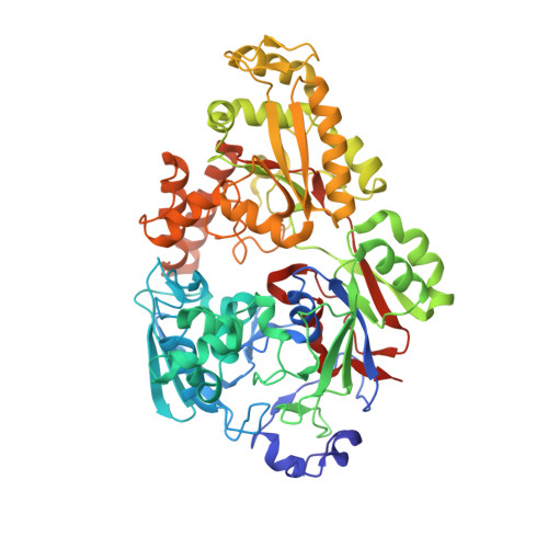
    Gram-negative Sphingomonas sp. strain A1 exhibits positive chemotaxis toward acidic polysaccharide pectin. SPH1118 has been identified as a pectin-binding protein involved in both pectin chemotaxis and assimilation. Here we show tertiary structures of SPH1118 with six different conformations as determined by X-ray crystallography. SPH1118 consisted of two domains with a large cleft between the domains and substrates bound to positively charged and aromatic residues in the cleft through hydrogen bond and stacking interactions. Substrate-free SPH1118 adopted three different conformations in the open form. On the other hand, the two domains were closed in substrate-bound form and the domain closure ratio was changed in response to the substrate size, suggesting that the conformational change upon binding to the substrate triggered the expression of pectin chemotaxis and assimilation. This study first clarified that the solute-binding protein with dual functions recognized the substrate through flexible conformational changes in response to the substrate size.


  • Organizational Affiliation

    Laboratory of Basic and Applied Molecular Biotechnology, Division of Food Science and Biotechnology, Graduate School of Agriculture, Kyoto University, Uji, Kyoto, 611-0011, Japan.


Macromolecules
Find similar proteins by:  (by identity cutoff)  |  3D Structure
Entity ID: 1
MoleculeChains Sequence LengthOrganismDetailsImage
SPH1118
A, B
619Sphingomonas sp. A1Mutation(s): 0 
Entity Groups  
Sequence Clusters30% Identity50% Identity70% Identity90% Identity95% Identity100% Identity
Sequence Annotations
Expand
  • Reference Sequence
Oligosaccharides

Help

Entity ID: 2
MoleculeChains Length2D Diagram Glycosylation3D Interactions
2,6-anhydro-3-deoxy-L-threo-hex-2-enonic acid-(1-4)-alpha-D-galactopyranuronic acid-(1-4)-alpha-D-galactopyranuronic acid
C, D
3N/A
Small Molecules
Ligands 3 Unique
IDChains Name / Formula / InChI Key2D Diagram3D Interactions
EPE
Query on EPE

Download Ideal Coordinates CCD File 
E [auth A],
H [auth B]
4-(2-HYDROXYETHYL)-1-PIPERAZINE ETHANESULFONIC ACID
C8 H18 N2 O4 S
JKMHFZQWWAIEOD-UHFFFAOYSA-N
TLA
Query on TLA

Download Ideal Coordinates CCD File 
G [auth A],
M [auth B]
L(+)-TARTARIC ACID
C4 H6 O6
FEWJPZIEWOKRBE-JCYAYHJZSA-N
GOL
Query on GOL

Download Ideal Coordinates CCD File 
F [auth A],
I [auth B],
J [auth B],
K [auth B],
L [auth B]
GLYCEROL
C3 H8 O3
PEDCQBHIVMGVHV-UHFFFAOYSA-N
Experimental Data & Validation

Experimental Data

  • Method: X-RAY DIFFRACTION
  • Resolution: 1.92 Å
  • R-Value Free: 0.232 
  • R-Value Work: 0.210 
  • Space Group: P 1 21 1
Unit Cell:
Length ( Å )Angle ( ˚ )
a = 57.687α = 90
b = 83.657β = 101.036
c = 148.521γ = 90
Software Package:
Software NamePurpose
REFMACrefinement
XDSdata reduction
XDSdata scaling
MOLREPphasing

Structure Validation

View Full Validation Report


Entry History & Funding Information

Deposition Data


Funding OrganizationLocationGrant Number
Japan Society for the Promotion of Science (JSPS)Japan--

Revision History  (Full details and data files)

  • Version 1.0: 2022-08-17
    Type: Initial release
  • Version 1.1: 2023-11-29
    Changes: Data collection, Refinement description