NASP maintains histone H3-H4 homeostasis through two distinct H3 binding modes.
Bao, H., Carraro, M., Flury, V., Liu, Y., Luo, M., Chen, L., Groth, A., Huang, H.(2022) Nucleic Acids Res 50: 5349-5368
- PubMed: 35489058 Search on PubMedSearch on PubMed Central
- DOI: https://doi.org/10.1093/nar/gkac303
- Primary Citation Related Structures:
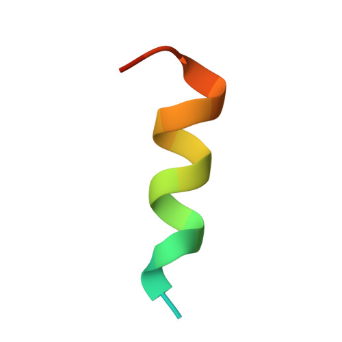
7V1K, 7V1L, 7V1M - PubMed Abstract:
Histone chaperones regulate all aspects of histone metabolism. NASP is a major histone chaperone for H3-H4 dimers critical for preventing histone degradation. Here, we identify two distinct histone binding modes of NASP and reveal how they cooperate to ensure histone H3-H4 supply. We determine the structures of a sNASP dimer, a complex of a sNASP dimer with two H3 α3 peptides, and the sNASP-H3-H4-ASF1b co-chaperone complex. This captures distinct functionalities of NASP and identifies two distinct binding modes involving the H3 α3 helix and the H3 αN region, respectively. Functional studies demonstrate the H3 αN-interaction represents the major binding mode of NASP in cells and shielding of the H3 αN region by NASP is essential in maintaining the H3-H4 histone soluble pool. In conclusion, our studies uncover the molecular basis of NASP as a major H3-H4 chaperone in guarding histone homeostasis.
- Key Laboratory of Molecular Design for Plant Cell Factory of Guangdong Higher Education Institutes, Department of Biology, School of Life Sciences, Southern University of Science and Technology, Shenzhen 518055, China.
Organizational Affiliation: