Structure of Klebsiella pneumoniae adenosine monophosphate nucleosidase.
Richardson, B.C., Shek, R., Van Voorhis, W.C., French, J.B.(2022) PLoS One 17: e0275023-e0275023
- PubMed: 36264993 Search on PubMedSearch on PubMed Central
- DOI: https://doi.org/10.1371/journal.pone.0275023
- Primary Citation Related Structures:
7UWQ - PubMed Abstract:
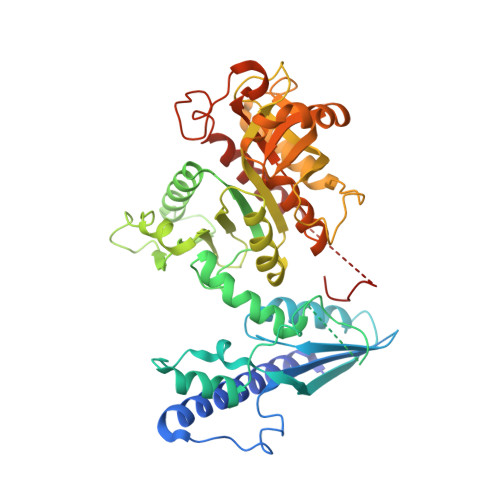
Klebsiella pneumoniae is a bacterial pathogen that is increasingly responsible for hospital-acquired pneumonia and sepsis. Progressive development of antibiotic resistance has led to higher mortality rates and creates a need for novel treatments. Because of the essential role that nucleotides play in many bacterial processes, enzymes involved in purine and pyrimidine metabolism and transport are ideal targets for the development of novel antibiotics. Herein we describe the structure of K. pneumoniae adenosine monophosphate nucleosidase (KpAmn), a purine salvage enzyme unique to bacteria, as determined by cryoelectron microscopy. The data detail a well conserved fold with a hexameric overall structure and clear density for the putative active site residues. Comparison to the crystal structures of homologous prokaryotic proteins confirms the presence of many of the conserved structural features of this protein yet reveals differences in distal loops in the absence of crystal contacts. This first cryo-EM structure of an Amn enzyme provides a basis for future structure-guided drug development and extends the accuracy of structural characterization of this family of proteins beyond this clinically relevant organism.
- The Hormel Institute, University of Minnesota, Austin, Minnesota, United States of America.
Organizational Affiliation: