7UVA

Crystal structure of KDM2A histone demethylase catalytic domain in complex with an H3C36 peptide modified by UNC8015


Experimental Data Snapshot

  • Method: X-RAY DIFFRACTION
  • Resolution: 1.98 Å
  • R-Value Free: 0.237 
  • R-Value Work: 0.183 
  • R-Value Observed: 0.186 

wwPDB Validation   3D Report Full Report


Ligand Structure Quality Assessment 


This is version 1.3 of the entry. See complete history


Literature

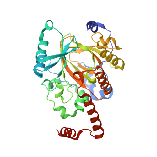
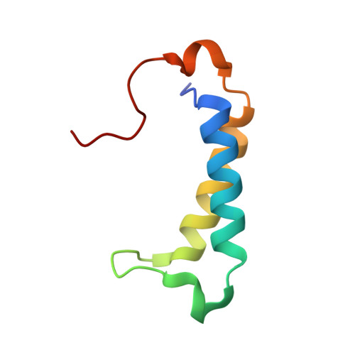
Structural basis of paralog-specific KDM2A/B nucleosome recognition.

Spangler, C.J.Skrajna, A.Foley, C.A.Nguyen, A.Budziszewski, G.R.Azzam, D.N.Arteaga, E.C.Simmons, H.C.Smith, C.B.Wesley, N.A.Wilkerson, E.M.McPherson, J.E.Kireev, D.James, L.I.Frye, S.V.Goldfarb, D.McGinty, R.K.

(2023) Nat Chem Biol 19: 624-632

  • DOI: https://doi.org/10.1038/s41589-023-01256-y
  • Primary Citation of Related Structures:  
    7UV9, 7UVA

  • PubMed Abstract: 

    The nucleosome acidic patch is a major interaction hub for chromatin, providing a platform for enzymes to dock and orient for nucleosome-targeted activities. To define the molecular basis of acidic patch recognition proteome wide, we performed an amino acid resolution acidic patch interactome screen. We discovered that the histone H3 lysine 36 (H3K36) demethylase KDM2A, but not its closely related paralog, KDM2B, requires the acidic patch for nucleosome binding. Despite fundamental roles in transcriptional repression in health and disease, the molecular mechanisms governing nucleosome substrate specificity of KDM2A/B, or any related JumonjiC (JmjC) domain lysine demethylase, remain unclear. We used a covalent conjugate between H3K36 and a demethylase inhibitor to solve cryogenic electron microscopy structures of KDM2A and KDM2B trapped in action on a nucleosome substrate. Our structures show that KDM2-nucleosome binding is paralog specific and facilitated by dynamic nucleosomal DNA unwrapping and histone charge shielding that mobilize the H3K36 sequence for demethylation.


  • Organizational Affiliation

    Department of Biochemistry and Biophysics, School of Medicine, University of North Carolina at Chapel Hill, Chapel Hill, NC, USA.


Macromolecules
Find similar proteins by:  (by identity cutoff)  |  3D Structure
Entity ID: 1
MoleculeChains Sequence LengthOrganismDetailsImage
Lysine-specific demethylase 2A
A, D
330Mus musculusMutation(s): 0 
Gene Names: Kdm2aFbl11Fbxl11Jhdm1aKiaa1004
EC: 1.14.11.27
UniProt
Find proteins for P59997 (Mus musculus)
Explore P59997 
Go to UniProtKB:  P59997
Entity Groups  
Sequence Clusters30% Identity50% Identity70% Identity90% Identity95% Identity100% Identity
UniProt GroupP59997
Sequence Annotations
Expand
  • Reference Sequence
Find similar proteins by:  (by identity cutoff)  |  3D Structure
Entity ID: 2
MoleculeChains Sequence LengthOrganismDetailsImage
Lysine-specific demethylase 2A
B, E
69Mus musculusMutation(s): 0 
Gene Names: Kdm2aFbl11Fbxl11Jhdm1aKiaa1004
EC: 1.14.11.27
UniProt
Find proteins for P59997 (Mus musculus)
Explore P59997 
Go to UniProtKB:  P59997
Entity Groups  
Sequence Clusters30% Identity50% Identity70% Identity90% Identity95% Identity100% Identity
UniProt GroupP59997
Sequence Annotations
Expand
  • Reference Sequence

Find similar proteins by:  Sequence   |   3D Structure  

Entity ID: 3
MoleculeChains Sequence LengthOrganismDetailsImage
Histone H3.2
C, F
13Homo sapiensMutation(s): 0 
UniProt & NIH Common Fund Data Resources
Find proteins for Q71DI3 (Homo sapiens)
Explore Q71DI3 
Go to UniProtKB:  Q71DI3
PHAROS:  Q71DI3
Entity Groups  
Sequence Clusters30% Identity50% Identity70% Identity90% Identity95% Identity100% Identity
UniProt GroupQ71DI3
Sequence Annotations
Expand
  • Reference Sequence
Experimental Data & Validation

Experimental Data

  • Method: X-RAY DIFFRACTION
  • Resolution: 1.98 Å
  • R-Value Free: 0.237 
  • R-Value Work: 0.183 
  • R-Value Observed: 0.186 
  • Space Group: P 21 21 21
Unit Cell:
Length ( Å )Angle ( ˚ )
a = 54.903α = 90
b = 87.054β = 90
c = 176.177γ = 90
Software Package:
Software NamePurpose
REFMACrefinement
XDSdata reduction
Aimlessdata scaling
PHASERphasing

Structure Validation

View Full Validation Report


Ligand Structure Quality Assessment 


Entry History & Funding Information

Deposition Data


Funding OrganizationLocationGrant Number
National Institutes of Health/National Institute of General Medical Sciences (NIH/NIGMS)United StatesT32GM119999
National Institutes of Health/National Institute of General Medical Sciences (NIH/NIGMS)United StatesR35GM133498
National Institutes of Health/National Cancer Institute (NIH/NCI)United StatesF99CA253730
National Institutes of Health/National Institute of General Medical Sciences (NIH/NIGMS)United StatesR35GM139514
National Institutes of Health/National Cancer Institute (NIH/NCI)United StatesR01CA010305

Revision History  (Full details and data files)

  • Version 1.0: 2023-02-22
    Type: Initial release
  • Version 1.1: 2023-03-01
    Changes: Database references
  • Version 1.2: 2023-05-17
    Changes: Database references
  • Version 1.3: 2023-10-25
    Changes: Data collection, Refinement description