7US3 | pdb_00007us3

Structure of Putrescine N-hydroxylase Involved Complexed with NADP+


Experimental Data Snapshot

  • Method: X-RAY DIFFRACTION
  • Resolution: 2.20 Å
  • R-Value Free: 
    0.223 (Depositor), 0.223 (DCC) 
  • R-Value Work: 
    0.182 (Depositor), 0.182 (DCC) 
  • R-Value Observed: 
    0.184 (Depositor) 

Starting Model: experimental
View more details

wwPDB Validation 3D Report Full Report

Validation slider image for 7US3

Ligand Structure Quality Assessment 


This is version 1.1 of the entry. See complete history

Literature

Kinetic and Structural Characterization of a Flavin-Dependent Putrescine N -Hydroxylase from Acinetobacter baumannii.

Lyons, N.S.Bogner, A.N.Tanner, J.J.Sobrado, P.

(2022) Biochemistry 61: 2607-2620

  • DOI: https://doi.org/10.1021/acs.biochem.2c00493
  • Primary Citation Related Structures: 
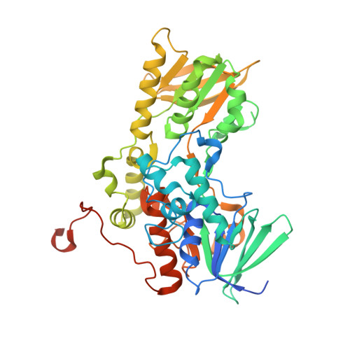
    7US3

  • PubMed Abstract: 

    Acinetobacter baumannii is a Gram-negative opportunistic pathogen that causes nosocomial infections, especially among immunocompromised individuals. The rise of multidrug resistant strains of A. baumannii has limited the use of standard antibiotics, highlighting a need for new drugs that exploit novel mechanisms of pathogenicity. Disrupting iron acquisition by inhibiting the biosynthesis of iron-chelating molecules (siderophores) secreted by the pathogen is a potential strategy for developing new antibiotics. Here we investigated FbsI, an N -hydroxylating monooxygenase involved in the biosynthesis of fimsbactin A, the major siderophore produced by A. baumannii. FbsI was characterized using steady-state and transient-state kinetics, spectroscopy, X-ray crystallography, and small-angle X-ray scattering. FbsI was found to catalyze the N -hydroxylation of the aliphatic diamines putrescine and cadaverine. Maximum coupling of the reductive and oxidative half-reactions occurs with putrescine, suggesting it is the preferred ( in vivo ) substrate. FbsI uses both NADPH and NADH as the reducing cofactor with a slight preference for NADPH. The crystal structure of FbsI complexed with NADP + was determined at 2.2 Å resolution. The structure exhibits the protein fold characteristic of Class B flavin-dependent monooxygenases. FbsI is most similar in 3D structure to the cadaverine N -hydroxylases DesB and DfoA. Small-angle X-ray scattering shows that FbsI forms a tetramer in solution like the N -hydroxylating monooxygenases of the SidA/IucD/PvdA family. A model of putrescine docked into the active site provides insight into substrate recognition. A mechanism for the catalytic cycle is proposed where dehydration of the C4a-hydroxyflavin intermediate is partially rate-limiting, and the hydroxylated putrescine product is released before NADP + .


  • Organizational Affiliation
    • Department of Biochemistry and Center for Drug Discovery, Virginia Tech, Blacksburg, Virginia 24061, United States.

Macromolecule Content 

  • Total Structure Weight: 220.84 kDa 
  • Atom Count: 14,794 
  • Modeled Residue Count: 1,712 
  • Deposited Residue Count: 1,864 
  • Unique protein chains: 1

Macromolecules

Find similar proteins by:|  3D Structure
Entity ID: 1
MoleculeChains  Sequence LengthOrganismDetailsImage
Putrescine N-hydroxylase
A, B, C, D
466Acinetobacter baumanniiMutation(s): 0 
Gene Names: AUO97_01770
EC: 1.14.13.252
UniProt
Find proteins for P0DXB2 (Acinetobacter baumannii (strain ATCC 17978 / DSM 105126 / CIP 53.77 / LMG 1025 / NCDC KC755 / 5377))
Explore P0DXB2 
Go to UniProtKB:  P0DXB2
Entity Groups
Sequence Clusters30% Identity50% Identity70% Identity90% Identity95% Identity100% Identity
UniProt GroupP0DXB2
Sequence Annotations
Expand
Reference Sequence

Small Molecules

Ligands 4 Unique
IDChains Name / Formula / InChI Key2D Diagram3D Interactions
FAD
(Subject of Investigation/LOI)

Query on FAD


Download:Ideal Coordinates CCD File
E [auth A],
I [auth B],
L [auth C],
O [auth D]
FLAVIN-ADENINE DINUCLEOTIDE
C27 H33 N9 O15 P2
VWWQXMAJTJZDQX-UYBVJOGSSA-N
NAP
(Subject of Investigation/LOI)

Query on NAP


Download:Ideal Coordinates CCD File
F [auth A],
H [auth B],
K [auth C],
N [auth D]
NADP NICOTINAMIDE-ADENINE-DINUCLEOTIDE PHOSPHATE
C21 H28 N7 O17 P3
XJLXINKUBYWONI-NNYOXOHSSA-N
PG4

Query on PG4


Download:Ideal Coordinates CCD File
J [auth C]TETRAETHYLENE GLYCOL
C8 H18 O5
UWHCKJMYHZGTIT-UHFFFAOYSA-N
PGE

Query on PGE


Download:Ideal Coordinates CCD File
G [auth A],
M [auth D]
TRIETHYLENE GLYCOL
C6 H14 O4
ZIBGPFATKBEMQZ-UHFFFAOYSA-N

Experimental Data & Validation

Experimental Data

  • Method: X-RAY DIFFRACTION
  • Resolution: 2.20 Å
  • R-Value Free:  0.223 (Depositor), 0.223 (DCC) 
  • R-Value Work:  0.182 (Depositor), 0.182 (DCC) 
  • R-Value Observed: 0.184 (Depositor) 
Space Group: P 21 21 21
Unit Cell:
Length ( Å )Angle ( ˚ )
a = 124.827α = 90
b = 126.314β = 90
c = 140.248γ = 90
Software Package:
Software NamePurpose
Aimlessdata scaling
PHENIXrefinement
PDB_EXTRACTdata extraction
XDSdata reduction
PHASERphasing

Structure Validation

View Full Validation Report


Ligand Structure Quality Assessment 


Entry History 

& Funding Information

Deposition Data


Funding OrganizationLocationGrant Number
National Science Foundation (NSF, United States)United StatesCHE-2003986
National Science Foundation (NSF, United States)United StatesCHE-2003658

Revision History  (Full details and data files)

  • Version 1.0: 2022-11-30
    Type: Initial release
  • Version 1.1: 2023-10-25
    Changes: Data collection, Refinement description