Hydrogens and hydrogen-bond networks in macromolecular MicroED data.
Clabbers, M.T.B., Martynowycz, M.W., Hattne, J., Gonen, T.(2022) J Struct Biol X 6: 100078-100078
- PubMed: 36507068 Search on PubMedSearch on PubMed Central
- DOI: https://doi.org/10.1016/j.yjsbx.2022.100078
- Primary Citation Related Structures:
7ULY - PubMed Abstract:
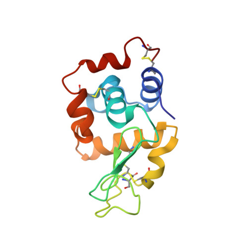
Microcrystal electron diffraction (MicroED) is a powerful technique utilizing electron cryo-microscopy (cryo-EM) for protein structure determination of crystalline samples too small for X-ray crystallography. Electrons interact with the electrostatic potential of the sample, which means that the scattered electrons carry information about the charged state of atoms and provide relatively stronger contrast for visualizing hydrogen atoms. Accurately identifying the positions of hydrogen atoms, and by extension the hydrogen bonding networks, is of importance for understanding protein structure and function, in particular for drug discovery. However, identification of individual hydrogen atom positions typically requires atomic resolution data, and has thus far remained elusive for macromolecular MicroED. Recently, we presented the ab initio structure of triclinic hen egg-white lysozyme at 0.87 Å resolution. The corresponding data were recorded under low exposure conditions using an electron-counting detector from thin crystalline lamellae. Here, using these subatomic resolution MicroED data, we identified over a third of all hydrogen atom positions based on strong difference peaks, and directly visualize hydrogen bonding interactions and the charged states of residues. Furthermore, we find that the hydrogen bond lengths are more accurately described by the inter-nuclei distances than the centers of mass of the corresponding electron clouds. We anticipate that MicroED, coupled with ongoing advances in data collection and refinement, can open further avenues for structural biology by uncovering the hydrogen atoms and hydrogen bonding interactions underlying protein structure and function.
- Department of Biological Chemistry, University of California, Los Angeles, CA 90095, United States.
Organizational Affiliation: