Download Mendeley
Structural Elucidation of a Protective B Cell Epitope on Outer Surface Protein C (OspC) of the Lyme Disease Spirochete, Borreliella burgdorferi.
Rudolph, M.J., Davis, S.A., Haque, H.M.E., Weis, D.D., Vance, D.J., Piazza, C.L., Ejemel, M., Cavacini, L., Wang, Y., Mbow, M.L., Gilmore, R.D., Mantis, N.J.(2023) mBio 14: e0298122-e0298122
- PubMed: 36976016 Search on PubMedSearch on PubMed Central
- DOI: https://doi.org/10.1128/mbio.02981-22
- Primary Citation Related Structures:
7UIJ, 7UJ2, 7UJ6 - PubMed Abstract:
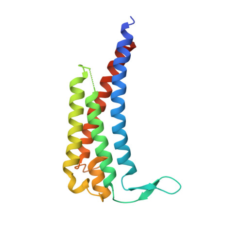
Outer surface protein C (OspC) plays a pivotal role in mediating tick-to-host transmission and infectivity of the Lyme disease spirochete, Borreliella burgdorferi. OspC is a helical-rich homodimer that interacts with tick salivary proteins, as well as components of the mammalian immune system. Several decades ago, it was shown that the OspC-specific monoclonal antibody, B5, was able to passively protect mice from experimental tick-transmitted infection by B. burgdorferi strain B31. However, B5's epitope has never been elucidated, despite widespread interest in OspC as a possible Lyme disease vaccine antigen. Here, we report the crystal structure of B5 antigen-binding fragments (Fabs) in complex with recombinant OspC type A (OspC A ). Each OspC monomer within the homodimer was bound by a single B5 Fab in a side-on orientation, with contact points along OspC's α-helix 1 and α-helix 6, as well as interactions with the loop between α-helices 5 and 6. In addition, B5's complementarity-determining region (CDR) H3 bridged the OspC-OspC' homodimer interface, revealing the quaternary nature of the protective epitope. To provide insight into the molecular basis of B5 serotype specificity, we solved the crystal structures of recombinant OspC types B and K and compared them to OspC A . This study represents the first structure of a protective B cell epitope on OspC and will aid in the rational design of OspC-based vaccines and therapeutics for Lyme disease. IMPORTANCE The spirochete Borreliella burgdorferi is a causative agent of Lyme disease, the most common tickborne disease in the United States. The spirochete is transmitted to humans during the course of a tick taking a bloodmeal. After B. burgdorferi is deposited into the skin of a human host, it replicates locally and spreads systemically, often resulting in clinical manifestations involving the central nervous system, joints, and/or heart. Antibodies directed against B. burgdorferi's outer surface protein C (OspC) are known to block tick-to-host transmission, as well as dissemination of the spirochete within a mammalian host. In this report, we reveal the first atomic structure of one such antibody in complex with OspC. Our results have implications for the design of a Lyme disease vaccine capable of interfering with multiple stages in B. burgdorferi infection.
- New York Structural Biology Center, New York, New York, USA.
Organizational Affiliation: