Chemically stable fluorescent proteins for advanced microscopy.
Campbell, B.C., Paez-Segala, M.G., Looger, L.L., Petsko, G.A., Liu, C.F.(2022) Nat Methods 19: 1612-1621
- PubMed: 36344833 Search on PubMedSearch on PubMed Central
- DOI: https://doi.org/10.1038/s41592-022-01660-7
- Primary Citation Related Structures:
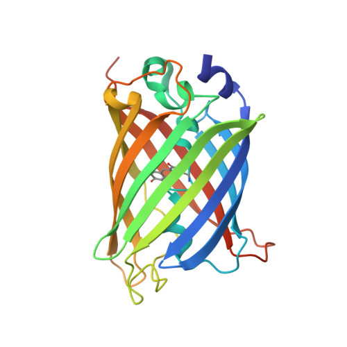
7UGR, 7UGS, 7UGT - PubMed Abstract:
We report the rational engineering of a remarkably stable yellow fluorescent protein (YFP), 'hyperfolder YFP' (hfYFP), that withstands chaotropic conditions that denature most biological structures within seconds, including superfolder green fluorescent protein (GFP). hfYFP contains no cysteines, is chloride insensitive and tolerates aldehyde and osmium tetroxide fixation better than common fluorescent proteins, enabling its use in expansion and electron microscopies. We solved crystal structures of hfYFP (to 1.7-Å resolution), a monomeric variant, monomeric hyperfolder YFP (1.6 Å) and an mGreenLantern mutant (1.2 Å), and then rationally engineered highly stable 405-nm-excitable GFPs, large Stokes shift (LSS) monomeric GFP (LSSmGFP) and LSSA12 from these structures. Lastly, we directly exploited the chemical stability of hfYFP and LSSmGFP by devising a fluorescence-assisted protein purification strategy enabling all steps of denaturing affinity chromatography to be visualized using ultraviolet or blue light. hfYFP and LSSmGFP represent a new generation of robustly stable fluorescent proteins developed for advanced biotechnological applications.
- Helen and Robert Appel Alzheimer's Disease Research Institute, Weill Cornell Medicine, New York, NY, USA. ben.campbell@protonmail.com.
Organizational Affiliation: