


Structure of the N-terminal domain of ViaA
Lemak, A., Reichheld, S., Bhandari, V., Houliston, S., Arrowsmith, C.H., Sharpe, S., Houry, W.A.To be published.
Experimental Data Snapshot
wwPDB Validation 3D Report Full Report

Entity ID: 1 | |||||
|---|---|---|---|---|---|
| Molecule | Chains | Sequence Length | Organism | Details | Image |
| Protein ViaA | 191 | Escherichia coli | Mutation(s): 0 Gene Names: viaA, GQW80_20505, GTP88_22555 | 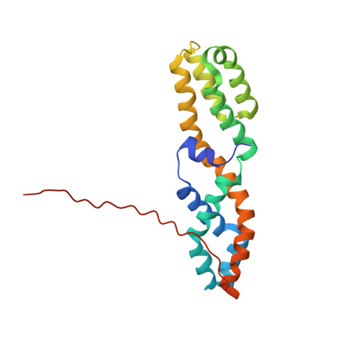 | |
UniProt | |||||
Entity Groups | |||||
| Sequence Clusters | 30% Identity50% Identity70% Identity90% Identity95% Identity100% Identity | ||||
| UniProt Group | P0ADN0 | ||||
Sequence AnnotationsExpand | |||||
Reference Sequence | |||||
| Funding Organization | Location | Grant Number |
|---|---|---|
| Canadian Institutes of Health Research (CIHR) | Canada | MOP-130374 |


