Identification of a novel deFADding activity in human, yeast and bacterial 5' to 3' exoribonucleases.
Sharma, S., Yang, J., Doamekpor, S.K., Grudizen-Nogalska, E., Tong, L., Kiledjian, M.(2022) Nucleic Acids Res 50: 8807-8817
- PubMed: 35904778 Search on PubMedSearch on PubMed Central
- DOI: https://doi.org/10.1093/nar/gkac617
- Primary Citation Related Structures:
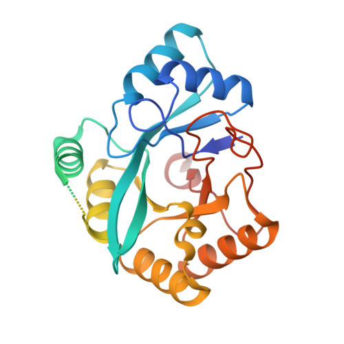
7UG9 - PubMed Abstract:
Identification of metabolite caps including FAD on the 5' end of RNA has uncovered a previously unforeseen intersection between cellular metabolism and gene expression. To understand the function of FAD caps in cellular physiology, we characterised the proteins interacting with FAD caps in budding yeast. Here we demonstrate that highly conserved 5'-3' exoribonucleases, Xrn1 and Rat1, physically interact with the RNA 5' FAD cap and both possess FAD cap decapping (deFADding) activity and subsequently degrade the resulting RNA. Xrn1 deFADding activity was also evident in human cells indicating its evolutionary conservation. Furthermore, we report that the recently identified bacterial 5'-3' exoribonuclease RNase AM also possesses deFADding activity that can degrade FAD-capped RNAs in vitro and in Escherichia coli cells. To gain a molecular understanding of the deFADding reaction, an RNase AM crystal structure with three manganese ions coordinated by a sulfate molecule and the active site amino acids was generated that provided details underlying hydrolysis of the FAD cap. Our findings reveal a general propensity for 5'-3' exoribonucleases to hydrolyse and degrade RNAs with 5' end noncanonical caps in addition to their well characterized 5' monophosphate RNA substrates indicating an intrinsic property of 5'-3' exoribonucleases.
- Department of Cell Biology and Neurosciences, Rutgers, University, Piscataway, NJ 08854, USA.
Organizational Affiliation: